
CASE HISTORY
A 23 years old female underwent bilateral percutaneous nephrostomy (PCN) placement for nephrolithiasis. Following the procedure, the patient developed progressive right-sided hemiparesis, which subsequently worsened with the development of altered sensorium.
Section Editors
Dr. Arpita Sahu, Dr. Sarbesh Tiwari, Dr. Sabha Ahmed,
Dr. Smily Sharma, Dr. Meena Nedunchelian
Authors
Dr. Genesis Giddo Koodaly — Fellow, Department of Neuroimaging and Interventional Radiology, NIMHANS, Bengaluru, India
Dr. Adarsh Mohapatra — Fellow, Department of Neuroimaging and Interventional Radiology, NIMHANS, Bengaluru, India
Dr. Meghana Kancharla, Fellow, Department of Neuroimaging and Interventional Radiology, NIMHANS, Bengaluru, India
Dr. Vignavishal Nandakumar – DM resident, Department of Neuroimaging and Interventional Radiology, NIMHANS, Bengaluru, India
Dr. Gauri Bhimrao Bornak — Senior Resident, Department of Neuroimaging and Interventional Radiology, NIMHANS, Bengaluru, India
Dr. Jitender Saini — Professor and Head, Department of Neuroimaging and Interventional Radiology, NIMHANS, Bengaluru, India
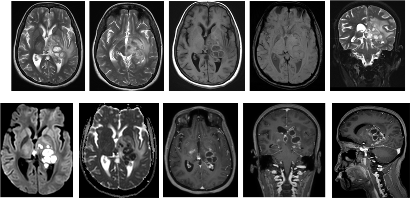
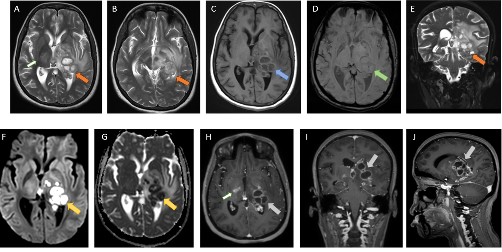
A–B: Axial T2; C: Axial T1; D: SWI; E: Coronal T2; F: DWI; G: ADC; H: Axial post-contrast T1; I: Coronal post-contrast T1; J: Sagittal post-contrast T1.
Multiple conglomerate and few discrete T2 hyperintense lesions are seen within the left thalamus and left medial temporal lobe, demonstrating a T2-intermediate signal intensity rim (orange arrows). The lesions show absent FLAIR suppression with both central and peripheral diffusion restriction (yellow arrows), resulting in expansion of the left thalamus. The lesion walls demonstrate intrinsic T1 hyperintensity (blue arrow). Patchy susceptibility blooming is seen on SWI (green arrow).
On post-contrast images, the lesions demonstrate peripheral enhancement with crenated margins (grey arrows). Few T2 hyperintense enhancing foci are seen in the right thalamic region (white arrows).
Significant perifocal vasogenic edema is seen extending into the left cerebral peduncle and hypothalamus, with resultant partial effacement of the left crural cistern and atrium of the left lateral ventricle. Mild upstream dilatation of the bilateral lateral ventricles is noted.
HISTOPATHOLOGY
Sections show small fragments of neuroparenchyma with a cyst wall thrown into folds, demonstrating extensive suppurative necrosis. The necrotic areas contain numerous fungal hyphae, which are long, slender, septate and branching, and are highlighted by Periodic acid–Schiff (PAS) and Grocott’s methenamine silver (GMS) stains.
The adjacent neuroparenchymal fragments show edema, along with chronic lymphocytic inflammation and scattered epithelioid histiocytes. Ziehl–Neelsen stain is negative for acid-fast bacilli.
Diagnosis: Necrotizing fungal inflammation (cerebral phaeohyphomycosis), consistent with Cladophialophora bantiana.
Cerebral phaeohyphomycosis is a rare but potentially fatal opportunistic fungal infection of the central nervous system caused by dematiaceous (melanized) fungi. Among these, Cladophialophora bantiana has a distinctive neurotropism and is an important cause of cerebral phaeohyphomycosis, particularly because infection may occur even in immunocompetent individuals. The route of CNS infection is predominantly hematogenous, with the lungs representing a potential portal of entry, although no primary extracranial focus may be demonstrable.
The imaging appearance is variable and may mimic other intracranial mass-forming lesions. Brain abscess formation is the most characteristic presentation, typically appearing as solitary or multiple lesions with surrounding vasogenic edema and variable rim enhancement. On MRI, the lesions are generally T2 hyperintense with a relatively hypointense or intermediate-signal capsule. Central diffusion restriction may be seen due to the high cellularity and viscous necrotic contents within the abscess cavity. Intrinsic T1 hyperintensity of the lesion wall, as observed in our case, may reflect the presence of melanin, proteinaceous contents, blood products, or a combination of these factors. Susceptibility effects, however, are variable and may be absent.
The presence of multiple conglomerate lesions with central diffusion restriction and a T2-intermediate rim suggested an infective abscess rather than a conventional neoplasm. The differential diagnosis of such lesions includes pyogenic abscess, tuberculoma, toxoplasmosis, metastases and high-grade glioma. Negative Ziehl–Neelsen staining in our case further argued against a mycobacterial etiology. Definitive diagnosis relies on histopathological examination and fungal identification.
The prognosis of cerebral C. bantiana infection remains poor, with high mortality despite antifungal therapy and surgical intervention. Early diagnosis, prompt surgical drainage or excision when feasible, and prolonged targeted antifungal therapy are important components of management.
Learning point: Cerebral phaeohyphomycosis should be considered in the differential diagnosis of rim-enhancing intracranial lesions with central diffusion restriction and disproportionate surrounding edema, even when the clinical history does not strongly suggest an invasive fungal infection.
References
1. Patil PN, Chavan R, Borse SS. Imaging in a rare case of cerebral phaeohyphomycosis caused by Cladophialophora bantiana in a renal transplant patient: a case report and the literature review. Egypt J Radiol Nucl Med. 2024;55:59. doi:10.1186/s43055-024-01229-8.
2. Rathod P, et al. Cerebral phaeohyphomycosis due to Cladophialophora bantiana in an immunocompetent individual: a case report and brief review of literature. Curr Med Mycol. 2020;6(2):52–57.
Add a Comment
You must be logged in to post a comment