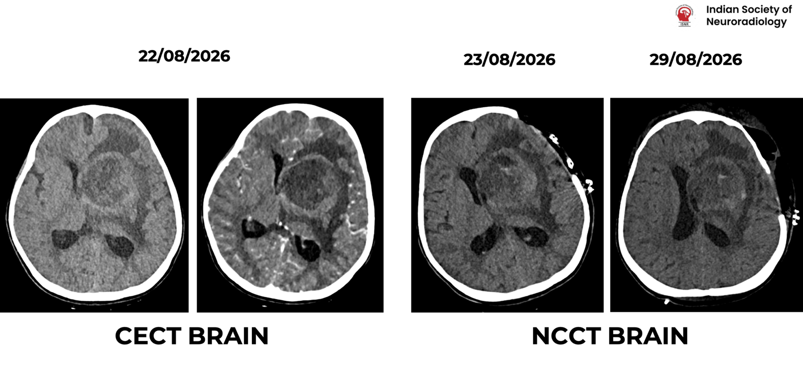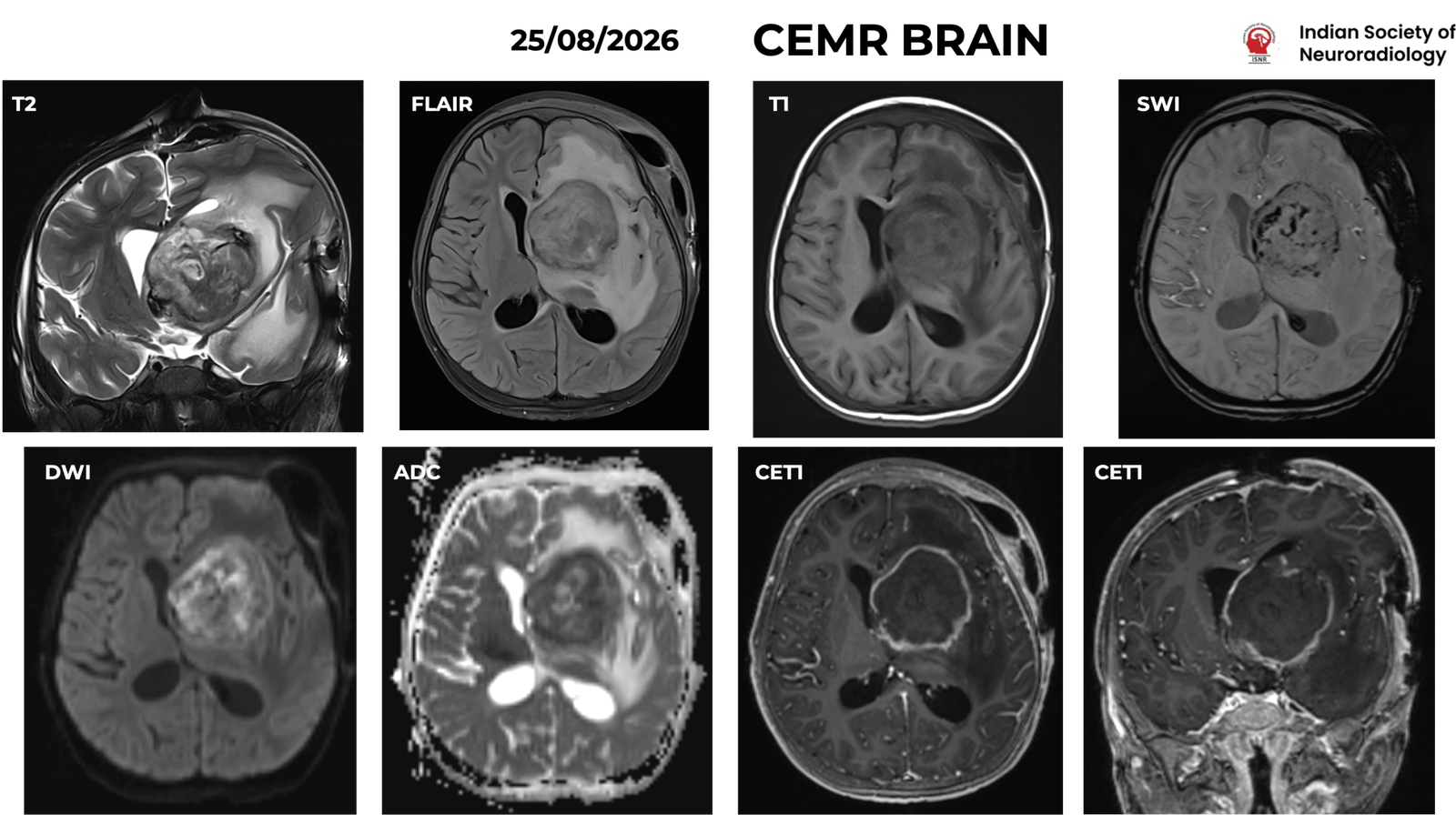
CASE HISTORY
• A 7-year-old girl was referred from IGICH with a 20-day history of headache, vomiting, and fever, followed by altered sensorium. Initial evaluation at a local hospital included lumbar puncture, which revealed hypoglycorrhachia (CSF glucose: 11 mg/dL) and elevated CSF protein (160 mg/dL). CSF culture grew Kocuria rosea, and antibiotic therapy was initiated.
• Her past history was significant for chronic diarrhea of 3 years’ duration and esophageal candidiasis. Serum IgE levels were elevated, raising the suspicion of an underlying hyper-IgE syndrome.
• Immunological profile: Markedly elevated serum IgE (~7,694 IU/mL), low serum IgM (<16 mg/dL), and absolute lymphocyte count of ~1,240 cells/µL, raising suspicion for an underlying immunodeficiency, including hyper-IgE syndrome.
• On examination, the child was conscious and oriented (GCS: E4V5M6). Both pupils were equal and reactive to light. Motor examination revealed reduced power in the right upper limb (3/5) and right lower limb (4+/5), with power greater than 3/5 in the left upper and lower limbs. Head circumference was 91.5 cm and mid-upper arm circumference (MUAC) was 13.5 cm. There was no history of loss of consciousness, seizures, or limb weakness reported in the initial history.
• Neuroimaging raised initial suspicion of an infective lesion. Antitubercular therapy (ATT) and dexamethasone were subsequently initiated.
• She underwent left frontal craniotomy with ultrasound-guided aspiration of the suspected abscess on 23/08/2026, followed by re-exploration and resection on 27/08/2026. In view of neurological deterioration, worsening cerebral edema, and midline shift, she underwent extension of the craniotomy to a left frontotemporoparietal decompressive craniectomy with right frontal external ventricular drain (EVD) placement on 29/08/2026.