
CASE HISTORY
Patient of relapsed Early T cell precursor Acute lymphoblastic leukemia ( BM + testis) . He underwent Matched unrelated donor hematopoietic stem cell transplantation. He relapsed in CNS 3 months post transplant and is on weekly triple IT for CNS relapse. Now presented with altered sensorium and slurring of speach.
Section Editors
Dr. Arpita Sahu, Dr. Sarbesh Tiwari, Dr. Sabha Ahmed,
Dr. Smily Sharma, Dr. Meena Nedunchelian
Authors
Dr Prakhyath Gmabhira D
DNB resident, KMCH, Coimbatore.
Dr Kannan G,
Consultant, KMCH, Coimbatore
Evaluation
Sepsis workup, cerebrospinal fluid analysis, and serum electrolytes were normal, prompting evaluation for structural cause.
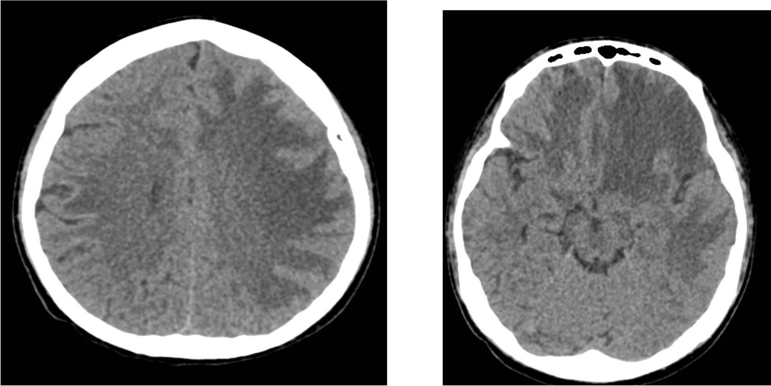
CT BRAIN:
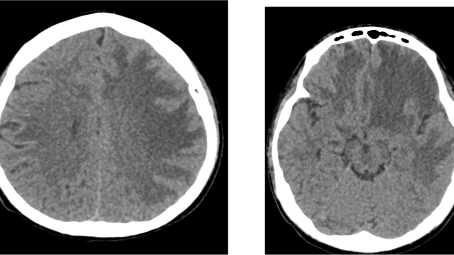
Bilateral confluent subcortical white matter hypodensities (left > right), involving fronto-parietal and temporal regions, with extension into the corpus callosum, associated with mild mass effect.
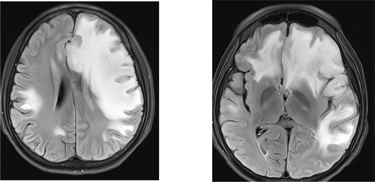
Diffuse infiltrative FLAIR hyperintense signal abnormalities involving:
o Bilateral fronto-parietal subcortical white matter (L > R)
o Anterior temporal subcortical white matter (L > R)
o Left lentiform nucleus and external capsule
o Periventricular white matter (trigone region)
o Bilateral basifrontal regions (L > R)
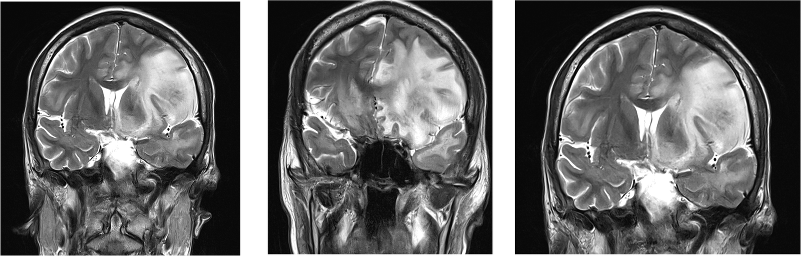
T2-weighted linear hypointense areas noted within the lesions.
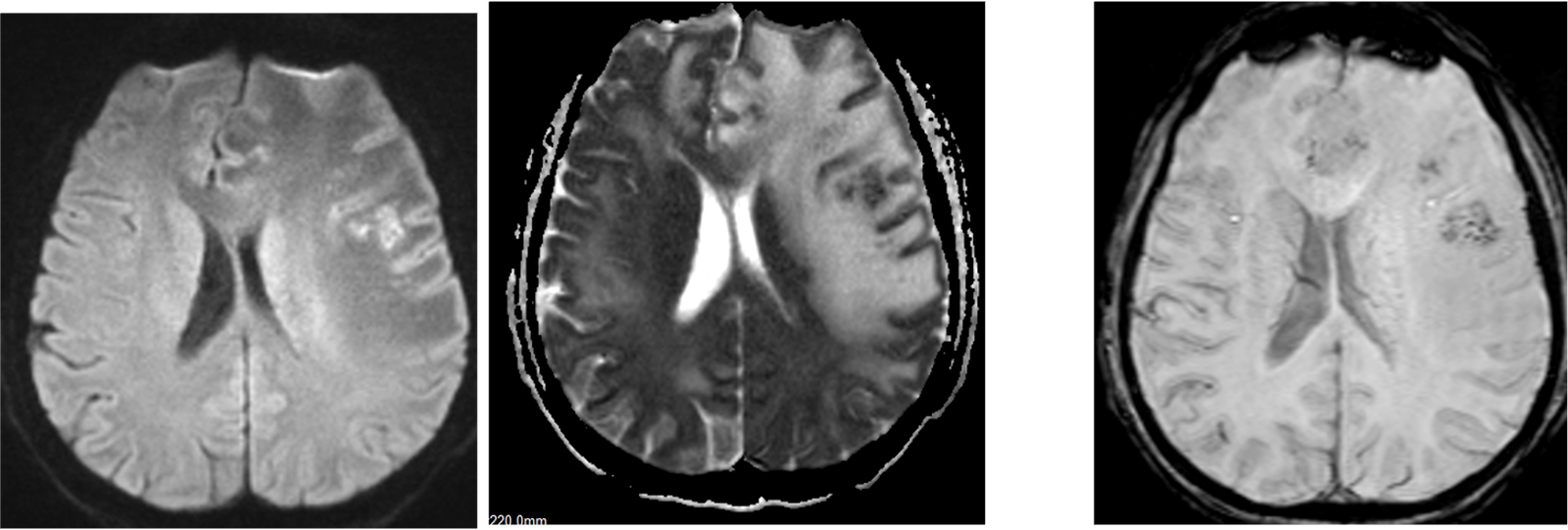
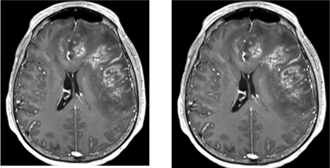
Patchy areas of diffusion restriction with associated blooming on SWI in the left frontal white matter.
Heterogeneous post-contrast enhancement involving the affected area are seen.
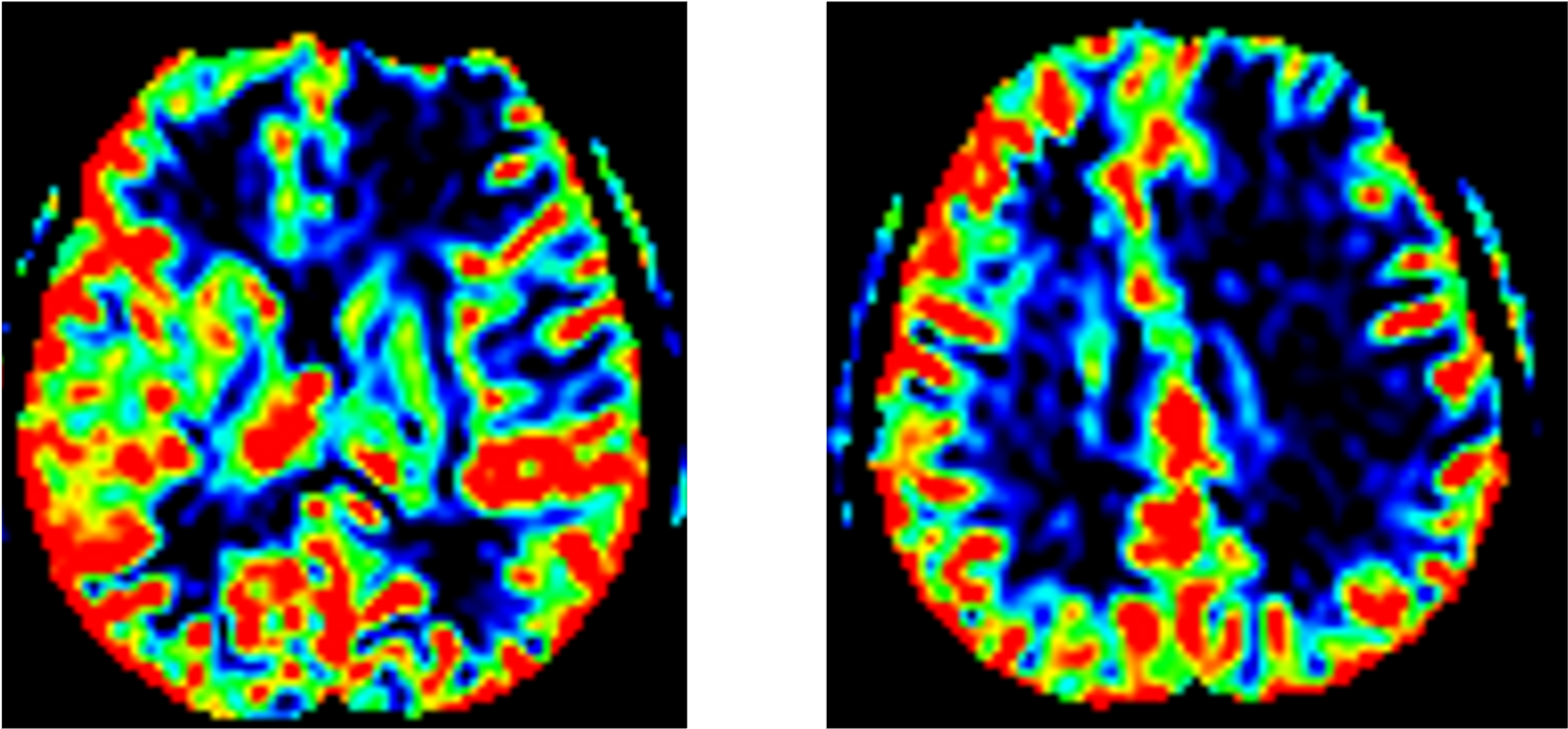
Significant reducedin cerebral blood volume (CBV) on perfusion imaging.
Imaging Differentials:
Angioinvasive fungal infection, likely invasive cerebral aspergillosis.
Other differentials- 1.leukemic infiltrates 2. PML
Diagnosis
High levels of Glactomannan in Bronchoalveolar lavage confirms Angio invasive cerebral aspergilosis

TREATMENT STARTED- INJECTION VARICONOZOLE.
After 1 week- SENSORIUM AND SPEECH IMPROVED.
ADVISE- CONTINUE ANTIFUNGAL.
Continued on voriconazole for 2 weeks and came for follow-up
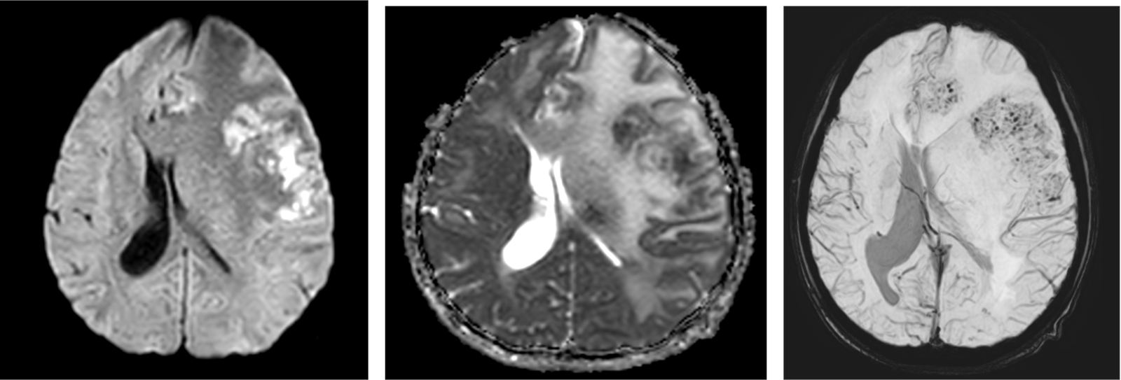
Interval increase in the patchy areas of diffusion restriction with peripheral SWI blooming are seen in left frontal lobe
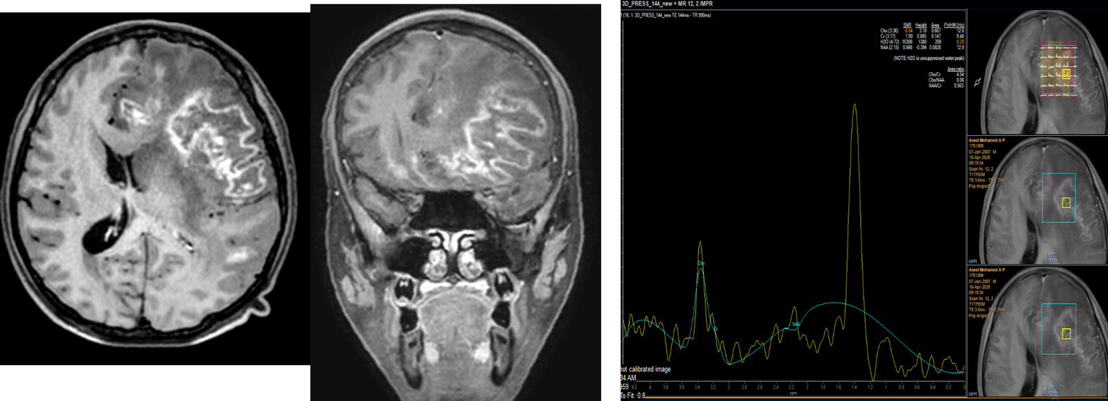
Peripheral enhancement are seen in left frontal lobe corresponding to areas of involvement, – likley evolving abscess. MRS shows a prominent lipid-lactate peak.
FURTHER TREATMENT
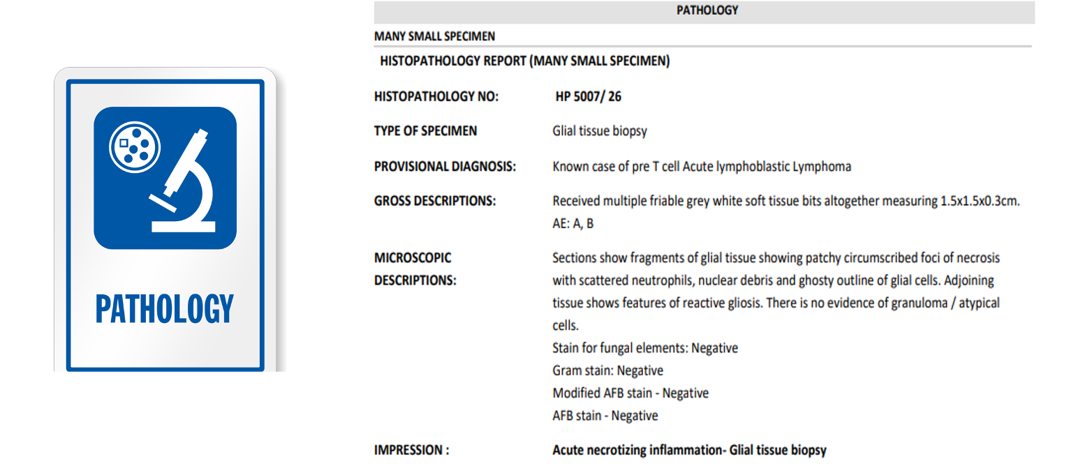
Left frontotemporal craniotomy & excision biopsy done. Antifungal treatment continued.
CONCLUSION:
| Feature | Angioinvasive Aspergillosis | CNS Leukemic Infiltrates | Progressive Multifocal Leukoencephalopathy |
| Pathophysiology | Angioinvasion causing vessel occlusion, infarction, and hemorrhage. | CNS relapse with proliferation of leukemic blasts. | JC virus opportunistic infection destroying oligodendrocytes (demyelination). |
| Primary Location | Corticomedullary junction, basal ganglia, thalami. | Leptomeninges, cranial nerves, parenchyma. | Asymmetric subcortical white matter (involves U-fibers). |
| Hemorrhage (SWI/T2)* | Prominent. Blooming artifact due to micro/macro-hemorrhages. | Typically absent. | Absent. |
| Diffusion (DWI/ADC) | Central restriction (due to ischemic/necrotic core). | Variable restriction (due to hypercellularity of blasts). | Usually absent (faint restriction possible at active advancing edge). |
| Enhancement (T1 C+) | Ring or heterogeneous enhancement. | Leptomeningeal or solid parenchymal enhancement. | Non-enhancing (unless IRIS develops). |
| T2/FLAIR Signal | “Target sign” (hyperintense core, dark rim, hyperintense edema). | Homogeneous hyperintensity. | Confluent, asymmetric hyperintensity. |
| T2 Internal Architecture | Linear/nodular hypointensities (represents fungal elements and blood products). | Homogeneous. Lacks dark internal bands. | Homogeneous. Lacks dark internal bands. |
| Lab Diagnostics | Elevated Galactomannan / Beta-D-glucan. | Positive CSF cytology / Flow cytometry for ALL blasts. | Positive JC Virus PCR in CSF. |
Discussion
●Post-HSCT/Immunocompromised status.
●T2-Weighted Clues: Linear hypointense areas noted within the infiltrative lesions—a classic sign of fungal hyphae or angioinvasive architecture.
●SWI (Susceptibility): Patchy areas of “blooming” in the left frontal white matter, indicating micro-hemorrhages.
●DWI (Diffusion): Patchy areas of restricted diffusion, suggesting acute infarction or high cellularity.
●Heterogeneous enhancement with mass effect.
●Perfusion Imaging: No significant increase in Cerebral Blood Volume (CBV), helping to differentiate from high-grade neoplastic processes.
REFERENCE:
1.Matis GK, Voultsinou D, Chrysou O, Birbilis T, Geroukis T. Cerebral aspergillosis and acute myeloid leukemia. J Neurosci Rural Pract. 2013 Aug;4(Suppl 1):S134-5. doi: 10.4103/0976-3147.116459. PMID: 24174784; PMCID: PMC3808046.
2.1.Machaj W, Korbecki A, Bińczyk W, Siudek B, Ussowicz M, Zimny A. Atypical radiological presentation of fatal invasive cerebral aspergillosis in a patient with acute lymphoblastic leukemia after allogeneic bone marrow transplant: A case report. Radiology Case Reports [Internet]. 2025 Jun 26;20(9):4590–3. Available from: https://www.sciencedirect.com/science/article/pii/S1930043325005266
3.Chantelot, L., Sitterlé, E., Poirée, S. et al. Cerebral Aspergillosis: Diagnostic Challenges, Therapeutic Strategies, and Future Research. CNS Drugs 40, 43–58 (2026). https://doi.org/10.1007/s40263-025-01241-0.