
CASE HISTORY
A 44-year-old woman with no previous significant medical history presented with a history of pain and electric shock like sensation on the right side of her face which aggravates on exposure to cold air for 4 years.
CASE CONTRIBUTED BY
Dr. Lakshmi Chaitanya, Department of Neuroimaging, Aster Whitefield Hospital, Bangalore
Dr. Sayani Mahal, Department of Neuroimaging, Aster Whitefield Hospital, Bangalore
Dr. Sunitha Palasamudram, Department of Neuroimaging, Aster Whitefield Hospital, Bangalore
Dr. Sathish Rudrappa, Department of Neurosurgery, Aster Whitefield Hospital, Bangalore
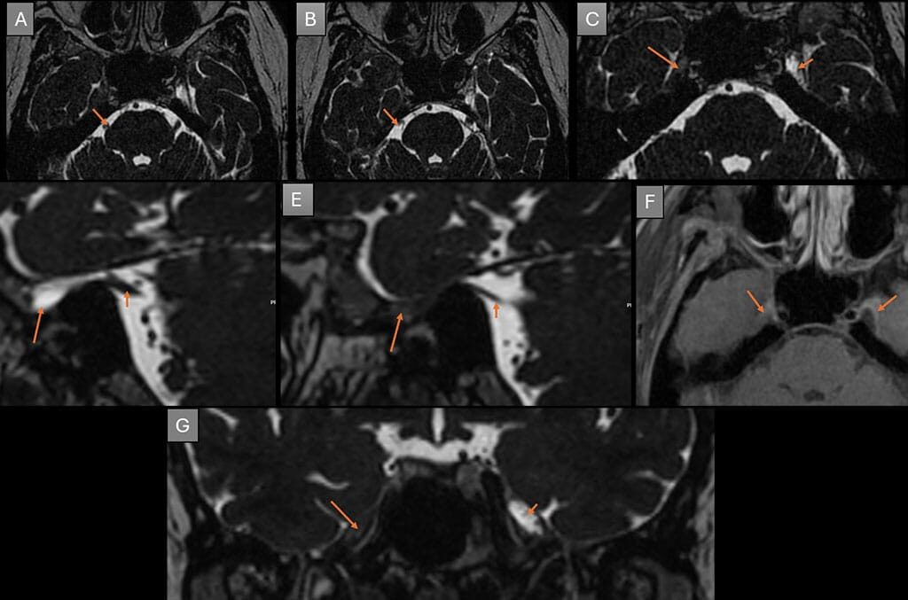
A – Axial heavily T2 weighted images showing normal root entry zone of right trigeminal nerve.
B -Axial heavily T2 weighted images showing atrophy of the right trigeminal nerve.
C – Axial heavily T2 weighted images shows absence of right Meckel’s cave (long arrow), normal Meckel’s cave on left side (short arrow).
D – Sagittal heavily T2 weighted image showing normal Meckel’s cave (long arrow) and trigeminal nerve (short arrow) on the left side.
E – Sagittal heavily T2 weighted image showing absent Meckel’s cave on the right side (long arrow) with relatively small caliber/ hypoplastic cisternal segment of trigeminal nerve (short arrow) compared to the left side.
F – Post contrast T1 weighted image enhancing dural sleeves of right Meckel’s cave. No other enhancing lesions along the course of right trigeminal nerve.
G – Coronal heavily T2 weighted image showing absent Meckel’s cave on right side (long arrow) and normal Meckel’s cave on left side (short arrow).
Diagnosis: ABSENT MECKELS CAVE ON THE RIGHT SIDE CAUSING TRIGEMINAL NEURALGIA
Trigeminal Neuralgia (TN):
Trigeminal neuralgia (TN) is a debilitating condition traditionally attributed to neurovascular conflict, where vascular structures compress the trigeminal nerve. However, in some patients, the absence of clear neurovascular conflict (NVC) on imaging necessitates a broader investigation into other potential causes. This review explores alternative etiologies of TN, specifically in cases where anatomical anomalies, including absent Meckel’s cave, are identified.
Other potential causes include tumors, multiple sclerosis, infection, trauma.
Prevalence: ~0.07%, more common in females.
High-resolution (3D T2 W) MRI is essential before any percutaneous trigeminal procedure to:
- Confirm the presence or absence of Meckel’s cave
- Identify alternative pathologies (e.g., NVC, tumor, demyelination, scarring)
Absence of Meckel’s Cave, as a rare etiology for TN:
- Meckel’s cave: a CSF-filled space in the middle cranial fossa housing the trigeminal ganglion.
- Its absence is extremely rare and has been linked to TN.
- Absence of CSF in the cave may cause ganglion injury leading to TN.
- Few reported cases worldwide, all in females (ages 35–80 years).
- The identification of an absent or obliterated Meckel’s cave on neuroimaging, although rare, can profoundly impact clinical decision-making and treatment planning.
REFERENCES:
- Jain A, Muneer MS, Okromelidze L, et al. Absence of Meckel cave: a rare cause of trigeminal neuralgia. AJNR Am J Neuroradiol. 2021;42(9):1610–1614.
- Lin J, Zhang Y, Li W, Yan J, Ke Y. Flatness of the Meckel cave may cause primary trigeminal neuralgia: a radiomics-based study. J Headache Pain. 2021;22(1):104.
- Shadani K, Kumar A, Rehan B, Banhwar IA. Absent Meckel’s cave in MRI, in a clinically diagnosed case of trigeminal neuralgia. A very rare case report. Pak J Radiol. 2020;30(4):293–295.
Add a Comment
You must be logged in to post a comment