
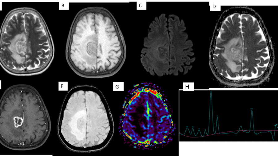
CASE HISTORY
• 53 year old female came with complaints of imbalance while standing and walking since 2 months. Patient was anaemic. No other co-morbidities. No significant past history.
• Patient underwent MRI brain imaging followed by whole body FDG PET-CT study.
Authors
1. Dr Shilali M S, Department of Radiology, Manipal Hospitals, Kanakapura Road, Bangalore.
2. Dr Lakshmikanth G N , Consultant, Department of Radiology, Manipal hospitals, Kanakapura Road, Bangalore.
3. Dr Chaitra P Adiga, Consultant, Department of Radiology, Manipal hospitals, Kanakapura Road, Bangalore.
4. Dr Yashwanth A N, Consultant, Department of Radiology, Manipal hospitals, Kanakapura Road, Bangalore.
5. Dr Sharath Kumar GG, HOD and Senior Consultant, Department of Radiology, Manipal Hospitals, Kanakapura Road, Bangalore.
DESCRIPTION
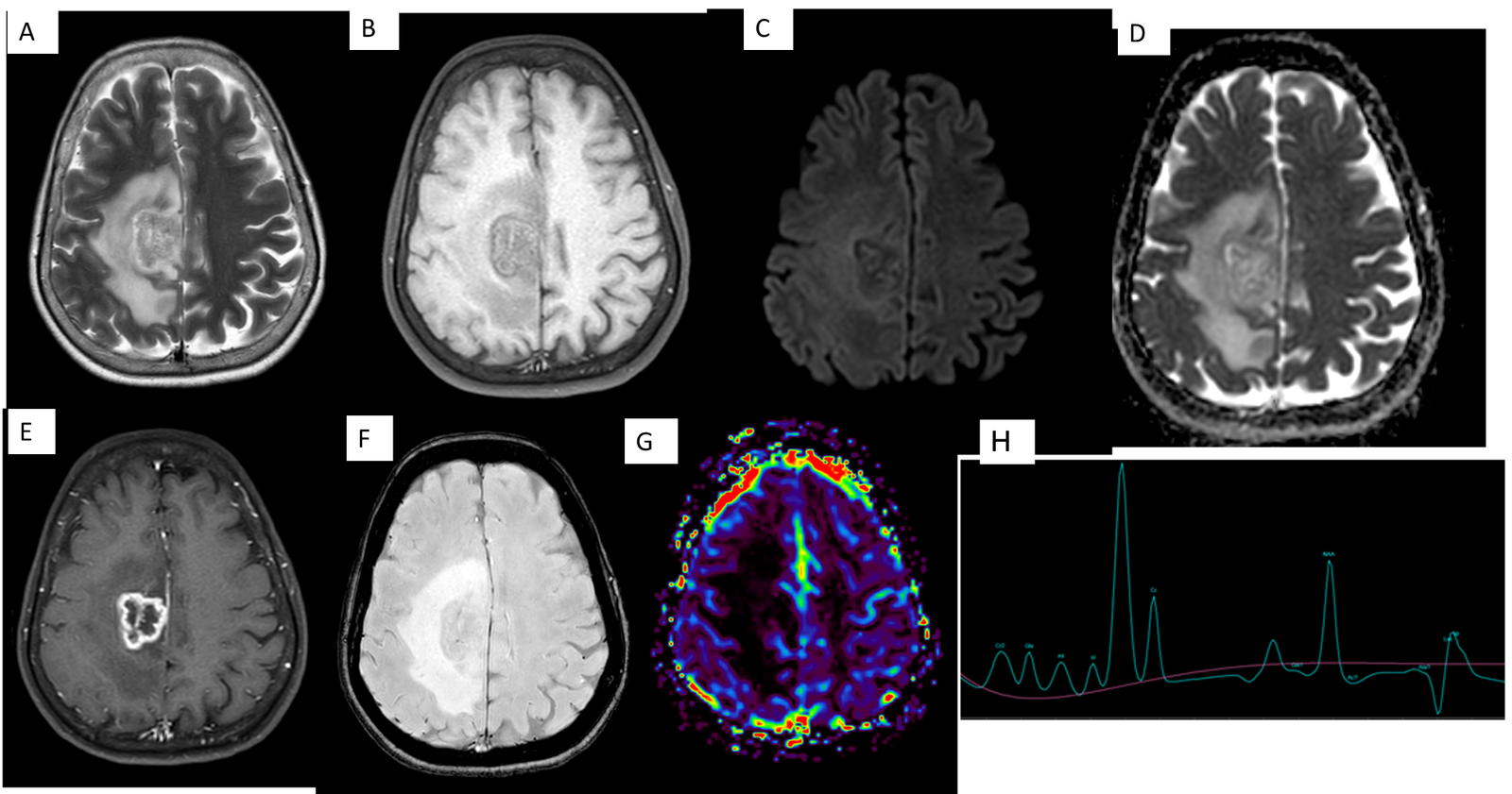
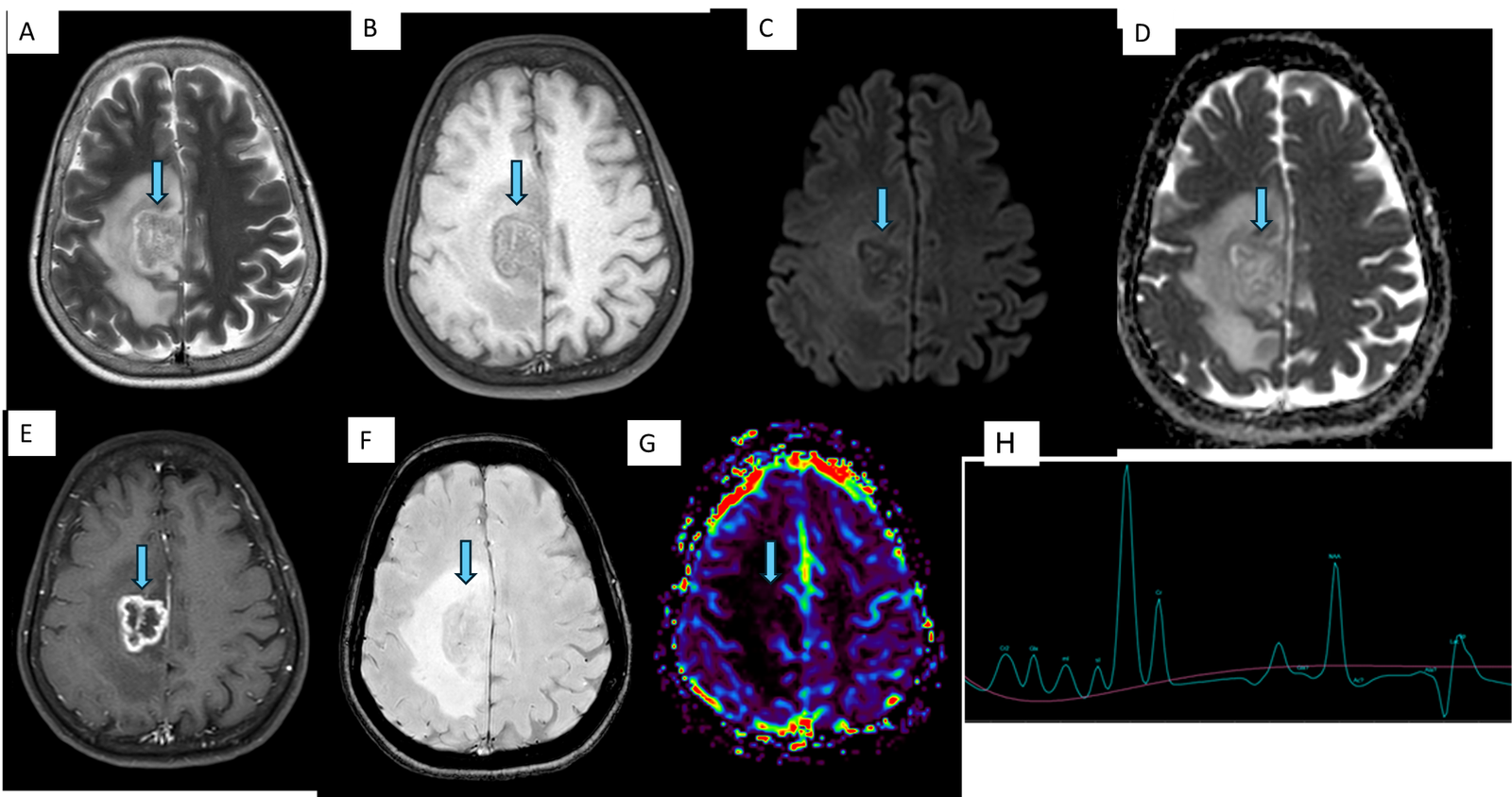
A & B – T2 and FLAIR axial images showing a well defined intra-axial heterogeneous and iso-hypointense lesion in the right high frontal lobe( mid cingulate gyrus ) with adjacent vasogenic edema.
C & D – DWI and ADC images showing no restricted diffusion.
E – Post contrast T1 images showing irregular rind of peripheral enhancement with central non enhancing necrotic areas. Mild enhancement of adjacent falx cerebri was also noted.
F : SWI images showing no areas of blooming.
G : Arterial spin labelling study showing no areas of hyper perfusion in the lesion or surrounding brain parenchyma
H : MR spectroscopy showing raised choline peak with raised choline to creatinine ratio. Lipid –lactate peak noted.
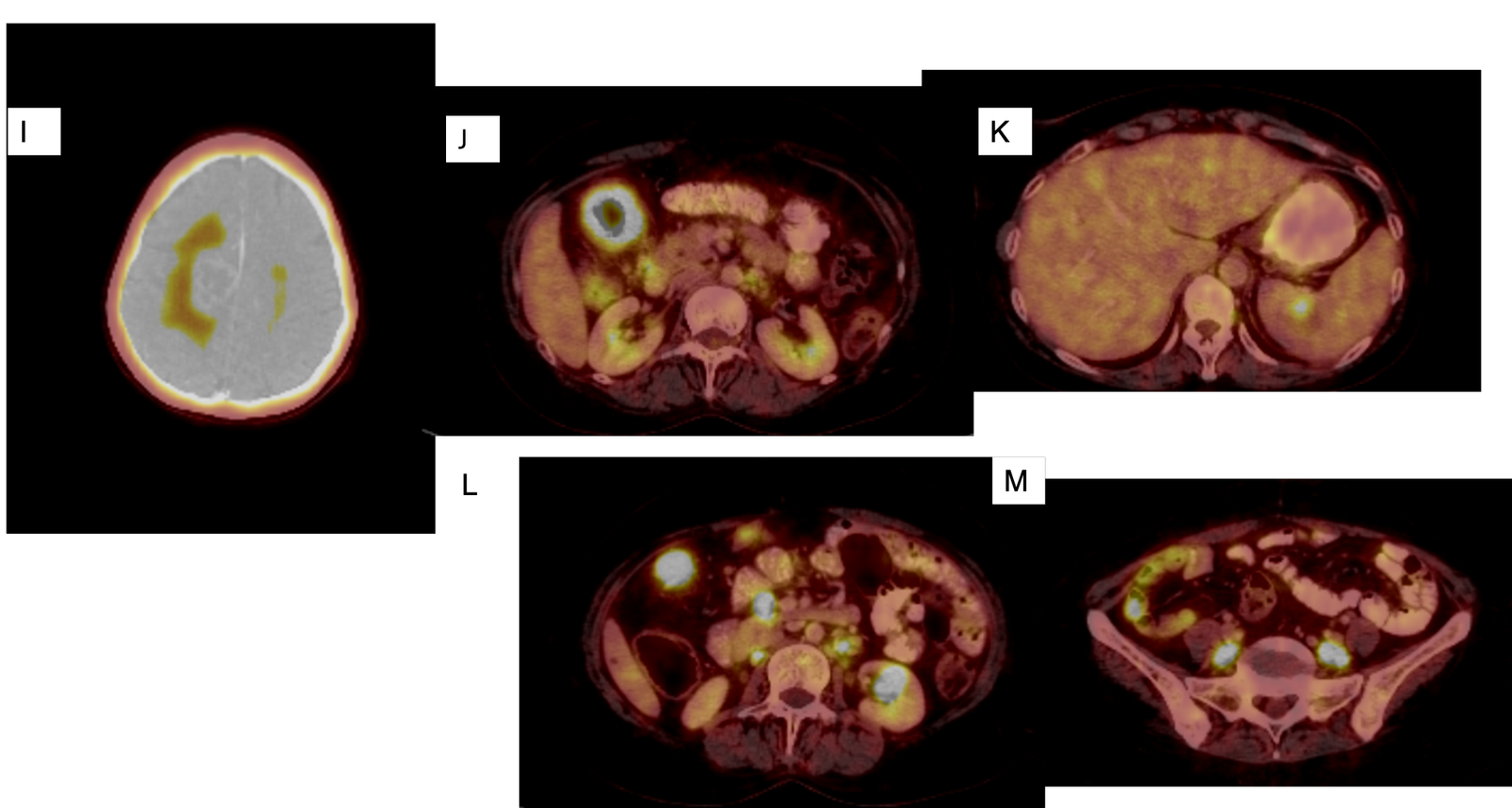
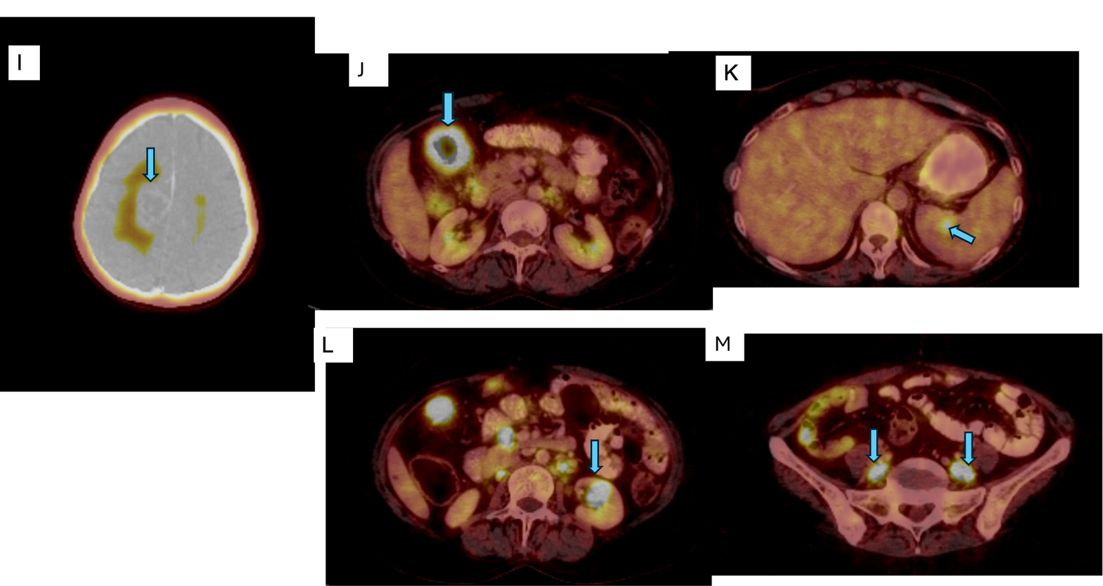
DESCRIPTION : 18F FDG PET-CT study
I – A tracer avid peripherally enhancing centrally necrotic mass lesion in the right high frontal lobe (mid cingulate gyrus) and adjacent vasogenic edema.
J – Tracer avid irregular enhancing circumferential wall thickening seen involving the hepatic flexure and proximal transverse colon.
K – Low grade tracer avid lesion in spleen.
L – Tracer avid lesion in the anterior cortex of the interpolar region of the left kidney.
M : Multiple tracer avid enlarged bilateral common iliac lymph nodes.
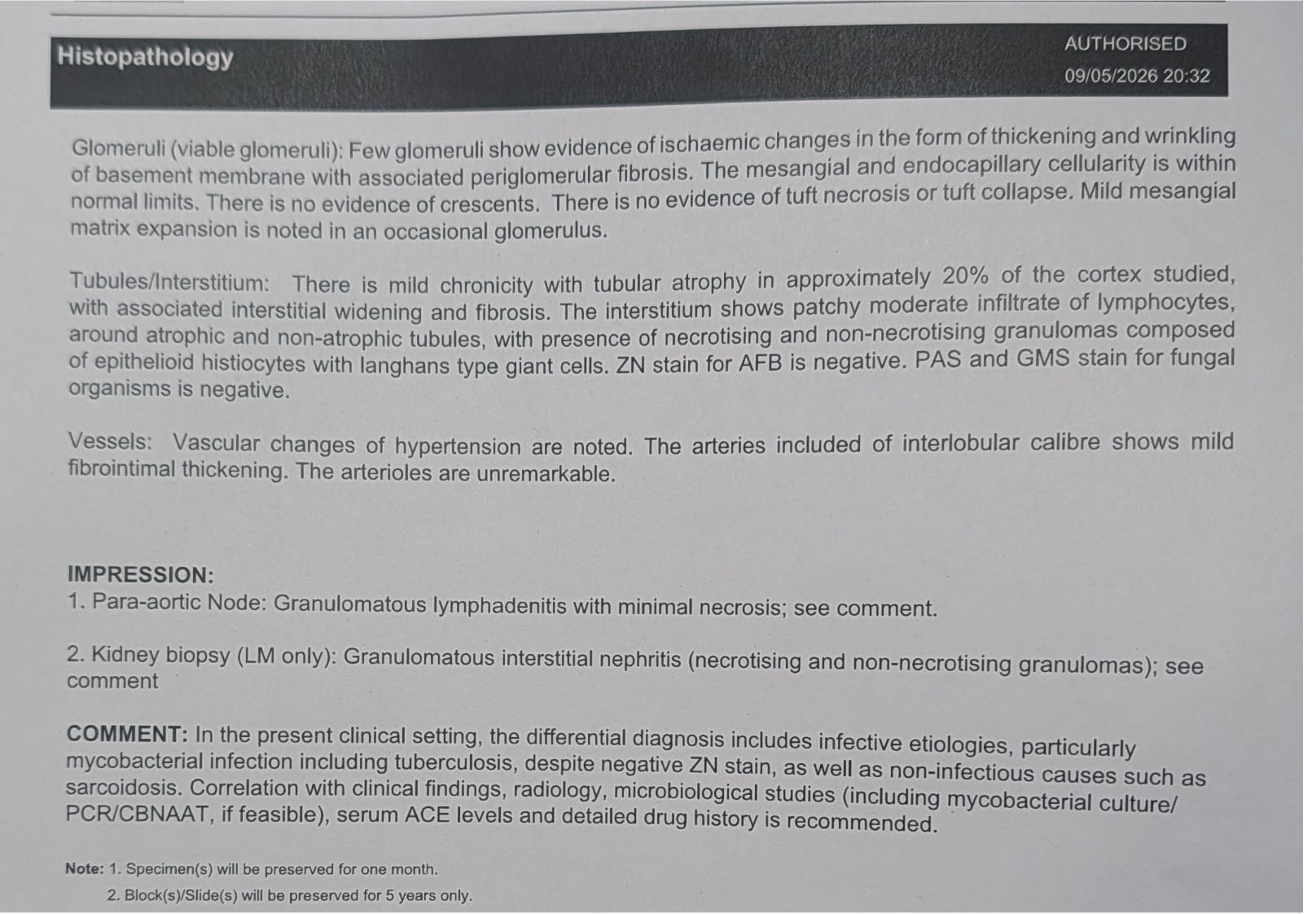
Final diagnosis: Disseminated Tuberculosis with cerebral tuberculoma
TUBERCULOMA –THE GREAT MASQUERADER
Discussion:
Background: Cerebral tuberculomas are granulomatous mass lesions resulting from hematogenous dissemination of Mycobacterium tuberculosis. On neuroimaging, tuberculomas can closely mimic primary or secondary intracranial neoplasms, particularly when presenting as solitary ring-enhancing lesions with surrounding vasogenic edema. Disseminated tuberculosis involving multiple organ systems may further complicate diagnosis by simulating metastatic malignancy on cross-sectional and metabolic imaging.
Key differentiating features
| Imaging Parameter | Tuberculoma | Metastasis |
| T2 signal | Often T2 hypointense (caseating granuloma) | Usually T2 hyperintense necrotic center |
| Lesion morphology | Conglomerate lesions common | Discrete lesions common |
| Meningeal enhancement | Relatively common | Uncommon |
| DWI | Variable | Usually no central restriction |
| Perfusion (rCBV) | Low | Increased |
| MR spectroscopy | Prominent lipid-lactate peak; lower Cho/Cr compared to aggressive tumors | Markedly elevated choline; higher Cho/Cr |
| 3.8 ppm peak on MRS | May be present | Rare |
| PET uptake | Can be intensely avid | Often avid |
| Ancillary findings | TB elsewhere, meningitis, tuberculomas | Known systemic malignancy |
In our case, the combination of a solitary necrotic ring-enhancing cerebral lesion with extensive perilesional edema and multiple FDG-avid extracranial lesions initially suggested metastatic disease. However, several features favoured tuberculosis, including absence of hyperperfusion on ASL, a prominent lipid–lactate peak on MR spectroscopy, lack of intralesional haemorrhage, mild adjacent falcine enhancement, and multisystem FDG-avid involvement compatible with disseminated tuberculosis. Histopathology ultimately confirmed the diagnosis.
Teaching point :
In endemic regions, a ring-enhancing lesion demonstrating low perfusion, T2 hypointense caseating components, and a dominant lipid-lactate peak should strongly raise suspicion for cerebral tuberculoma, even in the presence of multifocal FDG-avid systemic lesions suggestive of metastatic disease.
REFERENCES
• Koesbandono, Muljadi R, Sutanto R, et al. Intracranial tuberculomas: review of MRI findings and clinical features. Clinical Radiology. 2024;79(5):354-362. A recent comprehensive review of MRI characteristics of intracranial tuberculomas.
• Sankhe S, Baheti A, Ihare A, et al. Perfusion Magnetic Resonance Imaging Characteristics of Intracerebral Tuberculomas and Its Role in Differentiating Tuberculomas from Metastases. Acta Radiologica. 2013;54:307-312. Demonstrated significantly lower perfusion parameters in tuberculomas compared with metastases.
• Morales H, Alfaro D, Martinot C, et al. MR spectroscopy of intracranial tuberculomas: A singlet peak at 3.8 ppm as potential marker to differentiate them from malignant tumors. Neuroradiology Journal. 2015;28:294-302. Showed lower Cho/Cr ratios and a characteristic 3.8-ppm peak in tuberculomas compared with malignant tumors.
• Kumar V, Chawla S, Shah J, et al. Differentiation of tubercular infection and metastasis presenting as ring-enhancing lesions by diffusion and perfusion MRI. Journal of Neuroradiology. 2010;37:167-171. Demonstrated the value of combining diffusion and perfusion imaging in differentiating tuberculomas from metastases.