
November 2021
CASE HISTORY
INTERESTING CASE OF DELAYED AWAKENING FROM ANAESTHESIA POST SUPRATENTORIAL SURGERY
Case history
•An elderly woman presented with complaints of progressive diminution of vision in both eyes.
•MRI showed suprasellar meningioma impinging on the optic chiasm.
•Patient underwent excision of the mass .
•Post operatively the patient did not wake up from anaesthesia.
•CT scan was ordered to look for post-operative complications. • •
Case contributed by –
1.Dr Keerthinarayan M,
2.Dr.Vandana Peruri,
3.Dr.Hashim ,
4.Dr.Subodh,
5.Dr.Mythri,
Department of Radiology and Imaging ; Apollo Hospital, Sheshadripuram, Bengaluru.
 |  |
 |  |
 |  | 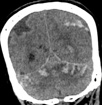 |
Immediate check CT head (Plain)
DIAGNOSIS?
 |  |
 |  |
 |  |  |
Remote cerebellar hemorrhage
| “Zebra sign” - Layering of blood along the cerebellar folia |  |
Remote cerebellar hemorrhage(RCH) is a rare but benign complication of the supratentorial and spine surgeries 1. It is called remote as the hemorrhage occurs distant from the site of surgery.
Most patients are asymptomatic. Other symptoms are of RCH are decreased level of consciousness, motor deficits, gait ataxia, and prolonged awakening from anesthesia as in our case 1. Many risk factors have been associated with RCH such as hypertension, epilepsy, compromised coagulation status, excess CSF loss during and after surgery and old age2.
Mechanism of occurrence is not well understood however the following hypotheses are proposed:
(a). Supratentorial procedures that involve opening of cisterns or ventricular systems with patients in a supine position may lead to opening of cisterns and the ventricular system causing CSF hypovolemia resulting in cerebellar sagging. This may cause transient occlusion of superior bridging veins of the posterior fossa leading to subsequent hemorrhagic infarction1,2.
(b). In contrast, some have theorized that increased pressure gradients across veins generated after removal of space occupying lesions such as tumors may lead to RCH 3.
REFERENCES
1. Amini A, Osborn AG, Mccall TD et-al. Remote cerebellar hemorrhage. Americal Journal of Neuroradiology. 2006;27 (2): 387-90.
2. Omer A, Engelman E, Bath K, Krauthamer A, Pisinski L. Remote cerebellar hemorrhage: A case report. Radiology Case Reports. 2019;14(3):385-389.
3. Konig A., Laas R., Herrmann H.D. Cerebellar haemorrhage as a complication after supratentorial craniotomy. Acta Neurochirurgica. 1987;88(3-4):104–108
Remote cerebellar hematoma
Remote cerebellar hemorrhage due to hemorrhagic venous infarction
Remote cerebellar hematoma