
Nov 2022
CASE HISTORY:
•11 year old female child presented with history of right sided hearing loss, vertigo and imbalance since 6 months, right facial numbness, facial asymmetry and difficulty swallowing since 3 months, headache and vomiting since 1 month duration.
•On neurological examination, there was involvement of the 5th,7th, 8th and lower cranial nerves on the right side.
•Tuning fork tests showed right SNHL. Cerebellar signs were present.
•Fundus examination showed papilledema suggestive of raised ICP.
•Working clinical diagnosis of right CP angle syndrome with increased ICP was made and imaging was performed.
•Case contributed by –
Dr Akshaya, Senior Resident, Department of Neuroimaging and Interventional Radiology, NIMHANS Bangalore.
Dr Jitender Saini, Professor, Department of Neuroimaging and Interventional Radiology, NIMHANS Bangalore
 |  |  |  | |
 |  |  |  |
 |  |  |  | |
 |  |  |  |
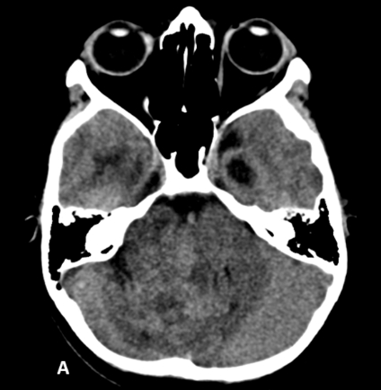
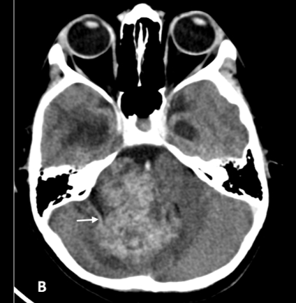
•Figure A & B: Axial CT sections before (A) and after (B) contrast administration show a large relatively well-defined heterogeneously enhancing lesion centered in the region of right cerebello-pontine cistern causing compression of the brainstem and fourth ventricle.
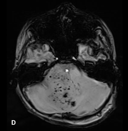
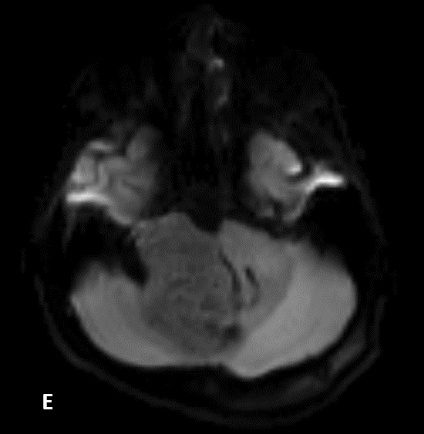
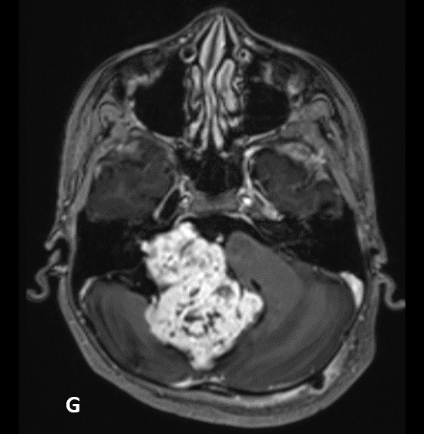
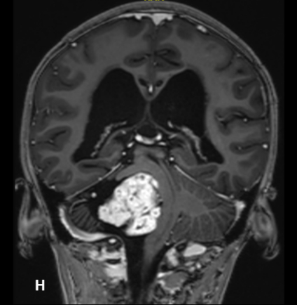
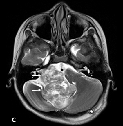
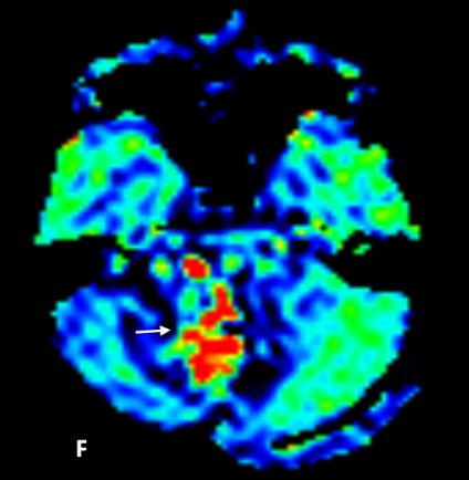
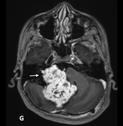
•Figures C to H: MRI images show T2 heterogeneous hyperintense lesion centered in the right cerebellopontine angle with intracanalicular extension (C), the lesion showing multiple susceptibility foci within on SWI (D), with no evidence of diffusion restriction (E). ASL CBF maps show areas of hypervascularity within the lesion (F). Post contrast images in axial (G) and coronal (H) planes show heterogeneous enhancement of the lesion with compression of the right MCP, brainstem and fourth ventricle.
FINAL DIAGNOSIS : Sporadic pediatric Vestibular Schwannoma
Background:
-Vestibular schwannomas (VS) are benign, slow-growing tumors that originate from vestibular division of the eighth cranial nerve.
-They typically originate in the internal acoustic meatus and can grow into the cerebellopontine angle, causing brainstem compression.
– Sporadic unilateral vestibular schwannomas without any NF2 characteristics or family history, are extremely rare in the paediatric population.
Clinical presentation
– SNHL is the most common presenting symptom of Pediatric VS similar to its adult counterpart.
– Headaches, tinnitus, ataxia and cerebellar dysfunction and signs of elevated ICP are the other presenting symptoms.
– Pediatric VS with their average increased size at presentation are more likely to cause symptoms due to mass effect and increased ICP than their adult counterparts. – Facial palsy and trigeminal hypesthesia are other less common presenting symptoms.
Learning point
•VSs are rare in childhood and an association with NF2 is often observed.
•Sporadic pediatric VS are extremely rare.
•Pediatric VS are more aggressive and vascular than their adult counterparts.
•Pediatric vestibular schwannomas cause hydrocephalus, various CN and brain stem findings by reaching quite large sizes.
•MRI in hypervascular VS commonly shows more solid tumour, without tumoral cyst and significantly larger size than non-hypervascular VS.
•The surface of hypervascular VS consistently shows multiple flow voids representing large draining veins.
•A multi-stage surgical approach is commonly used to treat hypervascular VS with the option of pre-operative embolization.
•Careful preoperative clinical-radiologic planning and intraoperative neuromonitoring are of critical importance for determining the surgical strategy and producing successful clinical results.
REFERENCES
•Malina, G. E. K., Heiferman, D. M., Riedy, L. N., Szujewski, C. C., Rezaii, E. G., Leonetti, J. P., & Anderson, D. E. (2020). Pediatric vestibular schwannomas: case series and a systematic review with meta-analysis, Journal of Neurosurgery: Pediatrics PED, 26(3), 302-310.
•Tomita T, Grahovac G. Cerebellopontine angle tumors in infants and children. Childs Nerv Syst. 2015 Oct;31(10):1739-50. doi: 10.1007/s00381-015-2747-x. Epub 2015 Sep 9. PMID: 26351227; PMCID: PMC4564453.
•Yamakami I, Kobayashi E, Iwadate Y, Saeki N, Yamaura A. Hypervascular vestibular schwannomas. Surg Neurol. 2002 Feb;57(2):105-12. doi: 10.1016/s0090-3019(01)00664-4. PMID: 11904203.
•Teranishi, Yu MD,,; Kohno, Michihiro MD, PhD,; Sora, Shigeo MD; Sato, Hiroaki MD; Nagata, Osamu MD, PhD. Hypervascular Vestibular Schwannomas: Clinical Characteristics, Angiographical Classification, and Surgical Considerations. Operative Neurosurgery: September 2018 – Volume 15 – Issue 3 – p 251-261
Large right schwannoma with heterogenous enchancement & hemorrhages within the lesion.
CP angle Ependymoma
Jugular foramen Schwannoma
D/D Choroid plexuses papiloma at CP Angle cistern.