
June 2022
CASE HISTORY:
•Insidious onset of protrusion of right eyeball, which has increased over 1 month.
•History of trivial fall 8 months ago with no loss of consciousness.
Neurological Examination
•Right eye proptosis with prominent veins
•Right temporal bruit.
•No motor deficits. Power 5/5. Tone is normal.
Case contributed by –
• Dr. Abin Jose, Radiology, Manipal Hospitals, Bangalore.
• Dr. Ullas V Acharya , Radiology, Manipal Hospitals, Bangalore.
 |  |  |  |
 |  |  |  |
 |  |  |  |
 |  |  |  |
Legends
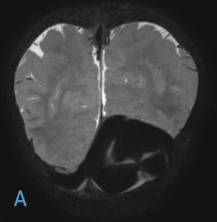
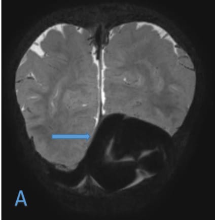
•A)Coronal T2W shows massively dilated proximal left transverse sinus possibly involving Torcula, suggestive of dural sinus malformation.
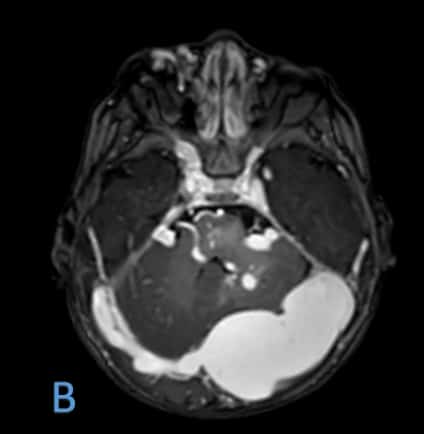
•B)Axial T1 PC shows contrast enhancement of the dural sinus malformation. No evidence of thrombosis within.
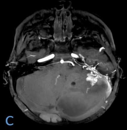
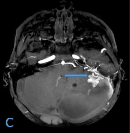
•C)Axial TOF MIP shows the fistula site to be located along the anterior, inferior and lateral aspects of the sac with prominent feeders from left internal carotid artery.
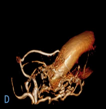
•D)Sagittal Volume rendered images show prominent arterial feeders from left middle meningeal artery and left occipital artery.
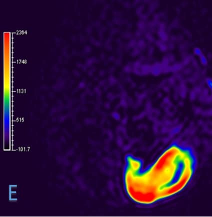
•E) Axial ASL sequence shows increased perfusion(arterial flow),suggestive of arteriovenous fistula.
•F) Coronal MRV MIP shows this dilated sac terminating as a prominent left sigmoid sinus with its continuity with the left internal jugular vein not being visualized, due to chronic occlusion.
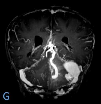
•G) Axial T1 PC shows pprominent posterior fossa veins.
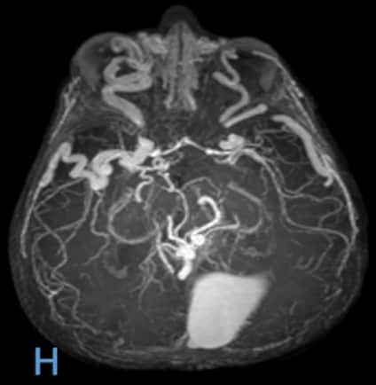
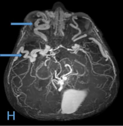
•H) Axial TOF MIP shows prominent bilateral superficial and deeper middle cerebral veins (right more than left) as well as dilated superior ophthalmic veins (right more than left) further draining across the canthi into supratrochlear veins been much more prominent in the right medial canthus. Also the dilated right superior ophthalmic vein is seen causing relative mass effect in the form of proptosis of the right eyeball.
Dural sinus malformation (DSM)
•Dural sinus malformation (DSM) is a rare congenital malformation which contains a dilated dural sinus pouch that communicates with the other sinuses and drains cerebral veins.
•They have been classified anatomically into a midline type and a lateral type, primarily based on the location.
• The midline type involves the posterior sinus with or without the torcula, with giant dural lakes and slow flow mural arteriovenous shunting, associated with spontaneous thrombosis, hemorrhagic infarction and a poorer prognosis.
•The lateral type involves the jugular bulb with otherwise normal sinuses and an associated high flow sigmoid sinus arteriovenous fistula. It has a good prognosis due to normal contralateral drainage pathway
Dural sinus malformation (DSM)
•Pathogenesis- Normal gestational remodelling of the ballooned sinuses is impeded by the high venous pressure due to the presence of dural arteriovenous fistulas.
•Another theory is that it results from excessive and disorganized development of the posterior sinuses, when they should be decreasing in size.
•References- •1. Fetal imaging of a rare case of dural sinus malformation: a case report. Kavya S. Kaushik, Ullas V. Acharya*, Rupa Ananthasivan, Bhavana Girishekar, Priyanka Kalidindi and Pooja G. Patil.
•2. Dural Sinus Malformations (DSM) with Giant Lakes, in Neonates and Infants. Review of 30 Consecutive Cases. M. Barbosa, J. Mahadevan.
Dural sinus malformation involving torcula with giant lakes
Turcular Dural sinus malformation with giant lakes
Dural sinus malformation
Dural-left transverse sinus Fistula with bilateral indirect CCFs
Vein of Galen malformation with aneurysmal dilatation
DSM