
July 2022
CASE HISTORY:
23 year old lady with tingling numbness in the right upper limb
No focal deficits
No significant past history
No neurological deficits on examination
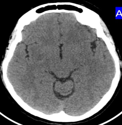
MRI BRAIN (P+C) done for further evaluation
Case contributed by –
Dr.Ganesh Shetty B
Dr.Lakshmikanth G N
Dr. Prakash P Iyer
Dr. Chaithra P Adiga
Department of Radiology, Apollo hospital, Sheshadripurum: Bangalore
 |  |  |  |
 |  |  |  |
 |  |  |  |
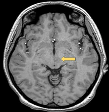 |  | 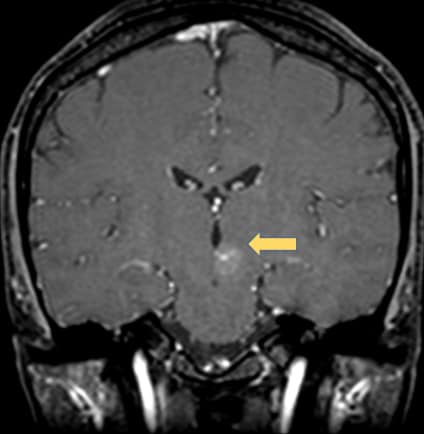 | 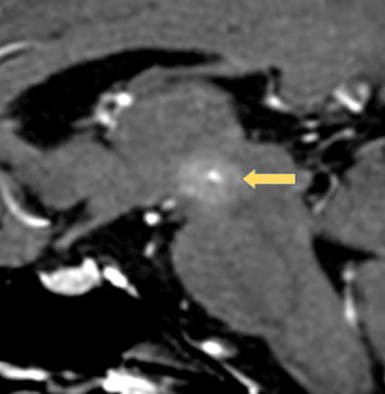 |
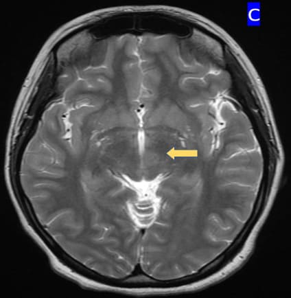
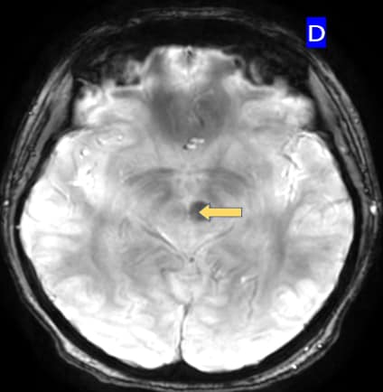
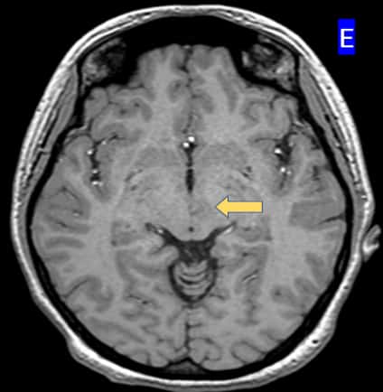
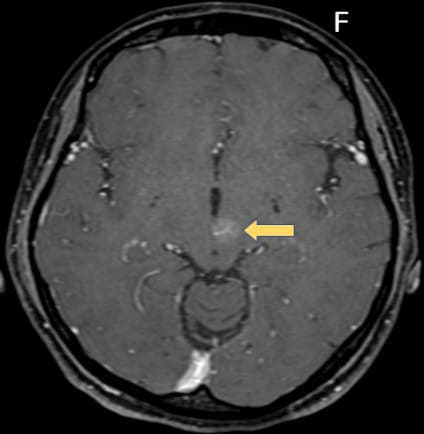
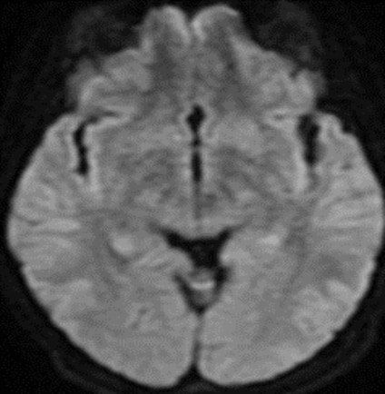
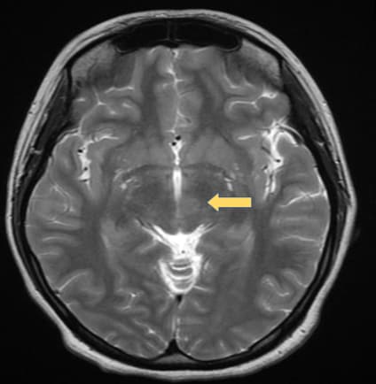
DIAGNOSIS: Capillary telangiectasias
•Mostly located in the brainstem (most commonly in the pons) (1)
•Often solitary and rarely can be multiple (1)
•Usually not seen on CT or catheter angiography (DSA)
•T1 may be iso to hypo and subtle bright on T2/FLAIR
•T2*GRE /SWI: Shows low signal intensity due to deoxyhemoglobin from sluggish flow and not due to bleed or mineralisation (2)
•Post contrast T1WI, may demonstrate stippled enhancement
•If large, can show branching/linear draining veins (3)
•No touch lesion & no follow-up is required if the imaging appearances are characteristic
REFERENCES
1.Castillo M, Morrison T, Shaw JA et-al. MR imaging and histologic features of capillary telangiectasia of the basal ganglia. AJNR Am J Neuroradiol. 2001;22 (8): 1553-5. AJNR Am J Neuroradiol
2.R R Lee et.al, Brain capillary telangiectasia: MR imaging appearance and clinicohistopathologic findings, December 1997 Radiology, 205, 797-805.
3.Auffray-Calvier E, Desal HA, Freund P, Laplaud D, Mathon G, de Kersaint-Gilly A. Capillary telangiectasias: angiographically occult vascular malformations—MRI symptomalogy apropos of 7 cases. J Neuroradiol 1999;26:257-261
Add a Comment
You must be logged in to post a comment