
February 2022
CASE HISTORY: 21 year old young male with H/O Global developmental delay with intellectual disability with seizure disorder and right eye retinal detachment.No history suggestive of perinatal ischemic insult.
Preliminary clinical suspicion :Mitochondrial vs Muscular dystrophy spectrum disorder
Investigation requested : MRI Brain
Case contributed by –
•Dr. Sabha Ahmed – Senior Resident, Dept Of Neuroimaging and Interventional Radiology, NIMHANS Bengaluru.
•Dr. Jitender Saini –Professor, Dept Of Neuroimaging and Interventional Radiology, NIMHANS Bengaluru.
•Dr. Vikram Holla- Assistant Professor, Dept Of Neurology, NIMHANS Bengaluru.
 |  |  | |
 |  |  |
 |  |  | |
 |  |  |
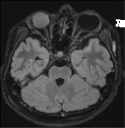
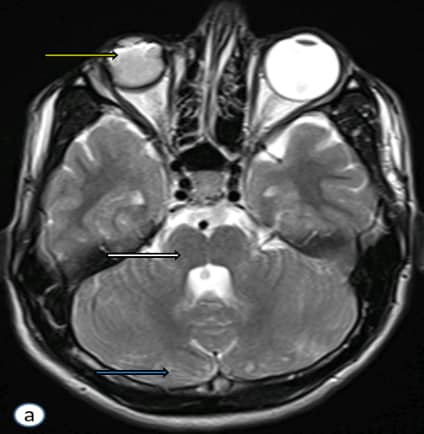
Axial T2 weighted image(a) show faint cystic changes showing FLAIR inversion(b) within the cerebellar parenchyma(blue arrow), pontine hypoplasia with a midline pontine cleft with absence of bulging contour of the facial colliculus(white arrow). Additionally phthisis bulbi of right eye observed(yellow arrow)
Axial T2 weighted sections at the level of medulla(c) show butterfly appearance of medulla oblongata
Axial sections of bone window(d) show undulating contour of the inner table of skull.
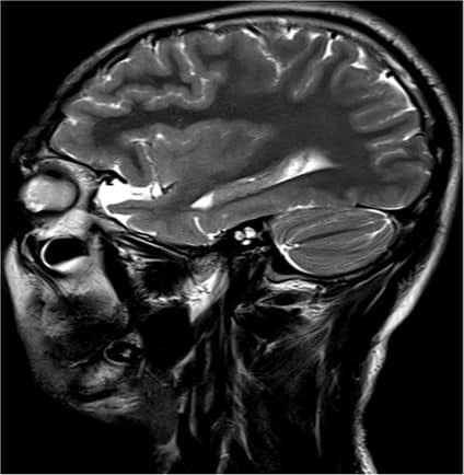
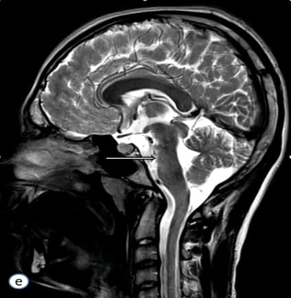
Sagittal T2(e) weighted sections at the level of massa intermedia show the profound brainstem hypoplasia.
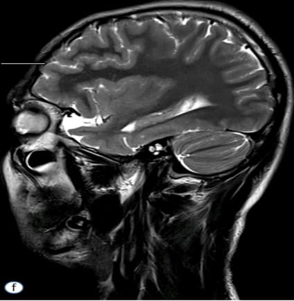
Parasagittal T2 (f)weighted sections at the level of temporal pole demonstrate polymicrogyric contour of the frontal cortical grey matter.
Muscle-Eye-Brain Disease Caused by Mutations in POMGNT1
•Genesis brought about by mutations in glycosylation of alpha-dystroglycan.
•Essentially , a “dystroglycanopathy’’ is used to describe a gamut of genetically heterogeneous disorders that include muscle, eye, and central nervous system involvement with a wide range of clinical severity(1)
•Manifestations range from mild limb girdle muscular dystrophy without cognitive impairment to severe syndromes with brain, eye- namely retinal, and muscle involvement(2)
•The medley of cortical dysplasia, brainstem hypoplasia, white matter signal changes, ventriculomegaly, and cerebellar cysts, may be representative of varied mutations in other alpha-dystroglycan related genes, however are more common in POMGnT1-mutated cases and should prompt tailored genetic investigations accordingly(3)
•Clinical learning :Muscle-eye-brain disease should be suspected in hypotonic infants presenting with generalized muscle weakness, elevated serum creatine kinase,phenotypic eye and brain malformations.
•Radiology learning : Constellation of brainstem abnormalities, cortical dysplasias andretinal changes should prompt a radiological suspicion of muscular dystrophies.
References
1.Muntoni F, Torelli S, Wells DJ, Brown SC. Muscular dystrophies due to glycosylation defects: diagnosis and therapeutic strategies. Curr Opin Neurol. 2011;24:437-442. 2.Godfrey C, Clement E, Mein R, et al. Refining genotype phenotype correlations in muscular dystrophies with defective glycosylation of dystroglycan. Brain. 2007;130:2725-2735. 3.Hehr U, Uyanik G, Gross C, et al. Novel POMGnT1 mutations define broader phenotypic spectrum of muscle-eye-brain disease. Neurogenetics. 2007;8:279-288. 28. Hu H, Candiello J, Zhang P, B
Add a Comment
You must be logged in to post a comment