
Feb 2023
CASE HISTORY:
•9 years old child presented to pediatrics emergency with cyanotic spell and headache. He was a known case of tetralogy of Fallot with a large VSD.
•On examination – the child had peripheral and central cyanosis.
•Child was treated with therapeutic phlebotomy and oxygen inhalation therapy. Cyanosis improved.
CASE CONTRIBUTED BY
Dr Rasi. Dr Khanak. Dr Poonam. Dr Sudhir Saxena.
Department of Diagnostic and Interventional Radiology. AIIMS. Rishikesh.
Dr Diksha Gupta. Department of Pediatrics. AIIMS. Rishikesh.
 |  |  |
 |  |
 |  |  |
 |  |
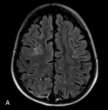
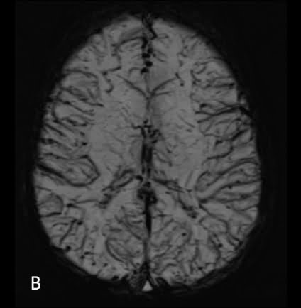
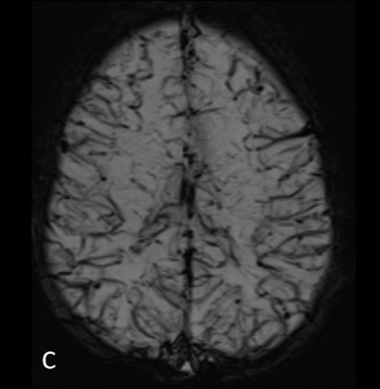
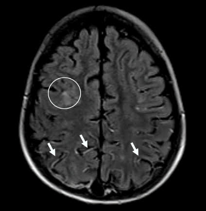
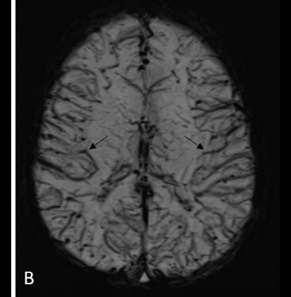
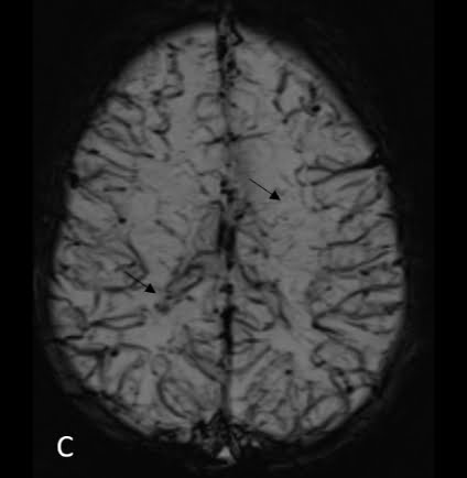
Images B &C – Axial Minimum intensity projection SWI images show distinctly congested, prominent and hypointense deeper and cortical veins (black arrows).
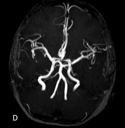
Image D – Axial MIP image of time-of-flight MR angiogram shows dilatation of bilateral ICA, MCA and vertebrobasilar system (black arrows).
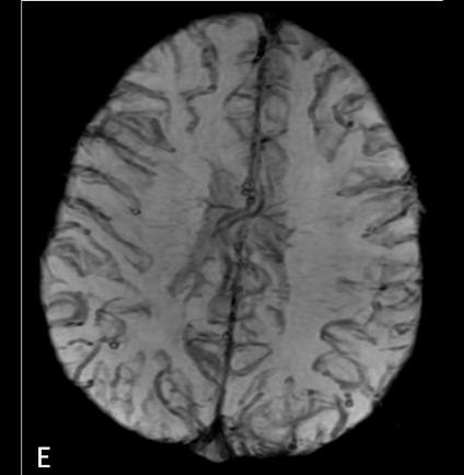
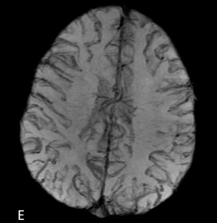
Image E – Axial Minimum intensity projection SWI image (post phlebotomy and inhalation oxygen therapy) shows partial reduction in venous congestion.
Diagnosis
Abnormal cerebral venous appearance and arterial dilatation due to systemic hypoxemia in tetralogy of Fallot and improvement following treatment.
Discussion
•Tetralogy of Fallot, a cyanotic heart diseases, causes arterio-venous blood mixing and resultant reduced blood oxygen saturation.
•Children with complex cyanotic heart diseases have hypoxic brain injury and delayed brain maturation. Small cerebral strokes, white matter intensities are associated with cyanotic heart diseases.
•Cerebral venous appearance varies according to blood oxygen saturation, deoxyhemoglobin levels and is a surrogate marker for hypoxic state (1). Improvement in blood oxygen saturation can improve cerebral venous congestion on SWI (2,3).
•Reduced cerebrovascular resistance results in arterial vasodilation. Inversely, hyperventilation and inhaled oxygen, in children who are scanned with general anesthesia, can result in higher venous signal on SWI (3).
References
•Verma RK, Keller D, Grunt S, Bigi S, Weisstanner C, Wiest R et al. Decreased oxygen saturation levels in neonates with transposition of great arteries: Impact on appearance of cerebral veins in susceptibility-weighted imaging. Scientific reports. 2017 Nov 13;7(1):15471.
•Mittal S, Wu Z, Neelavalli J, Haacke EM. Susceptibility-weighted imaging: technical aspects and clinical applications, part 2. American Journal of neuroradiology. 2009 Feb 1;30(2):232-52.
•Verschuuren S, Poretti A, Buerki S, Lequin MH, Huisman TA. Susceptibility-weighted imaging of the pediatric brain. American Journal of Roentgenology. 2012 May;198(5):W440-9.
Add a Comment
You must be logged in to post a comment