
December 2021
CASE HISTORY
•39 year man
•K/c/o of Type 2 DM
•Newly diagnosed microcytic anemia
•Progressive weakness of left upper limb and lower limb past 6 months
•Involuntary movements of left hand
Case contributed by –
Dr. Sadhana Beaty, Dr. Sai kanth Deepalam, Dr. Babu Phillip – Department of Radiodiagnosis & Intervention radiology St. Johns Medical college, Bangalore
 |  |  |
 |  |  |
 |  |  |
 |  |  |
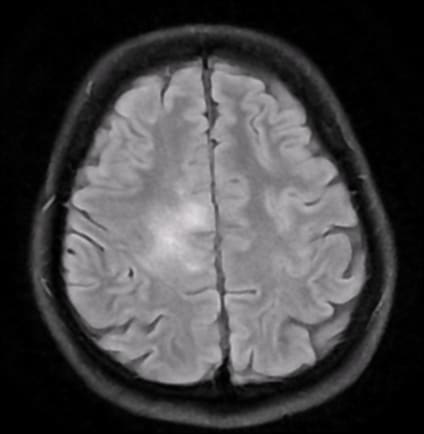
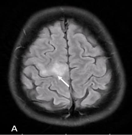
A and B: Axial T2 FLAIR
C : Coronal T2 FLAIR images show unilateral white matter hypointensity involving juxtacortical white matter in the pre central gyrus extending to the deep white matter (corticospinal tract)
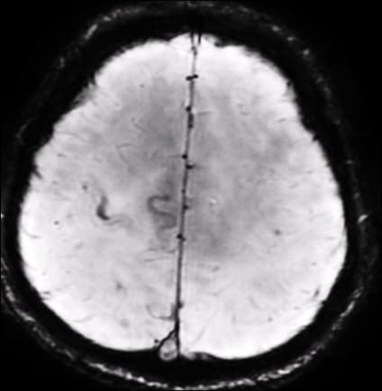
D : Axial SWI, Linear hypointensity is seen along the grey white matter junction on SWI
E : Axial T1 post contrast, shows no enhancement
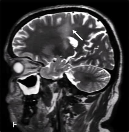
F: Sagittal T2WI, Punctate T2 hyperintensity is noted surrounding the main lesion resembling the ‘Milky way’
Progressive Multifocal Leukoencephalopathy (PML)
•PML is caused by reactivation of JC polymyovirus in immunocompromised patients with PML being the most common clinical manifestation of the infection (1,2).
•Small demyelinating lesions initially form in the subcortical U fibers, later progress into extensive white matter lesions involving deep white matter by fusion of small lesions and local expansion (1).
•JC virus spreads along the nerve tracts with the typical cerebral lesion extending from the corticomedullary border to the deep white matter with multiple punctate regions of high signal surrounding the main component (Milky Way sign)(1,2).
•Juxtacortical susceptibility changes corresponding to the FLAIR changes is due to iron accumulation in macrophages and is seen in long standing cases (2).
•Lesions in the posterior fossa can involve middle cerebellar peduncle and extend to cerebellar white matter draping the dentate nucleus (MRI Shrimp sign).
•Above case had low CD4 counts (Idiopathic CD4 lymphocytopenia). HIV was negative and CSF – JC virus is positive
REFERENCES
1. Ono D, Shishido-Hara Y, Mizutani S, Mori Y, Ichinose K, Watanabe M, et al. Development of demyelinating lesions in progressive multifocal leukoencephalopathy (PML): Comparison of magnetic resonance images and neuropathology of post-mortem brain. Neuropathology. 2019;39(4):294–306.
2. Mahajan KR, Amin M, Poturalski M, Lee J, Herman D, Zheng Y, et al. Juxtacortical susceptibility changes in progressive multifocal leukoencephalopathy at the gray–white matter junction correlates with iron-enriched macrophages. Mult Scler J. 2021;27(14):2159–69.
Add a Comment
You must be logged in to post a comment