Sept 2022
CASE HISTORY:
•A 30 year old man, presented with complaints of severe headache and vomiting since 4 days.
•No history of trauma. No focal neurological deficits.
•No significant past history.
Case contributed by –
Dr. Maria Varghese and Dr. Savith Kumar
Department of Radiology and Imaging sciences, Apollo hospitals, BG Road-Bangalore
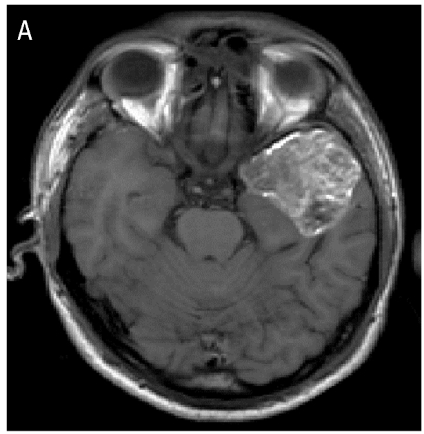
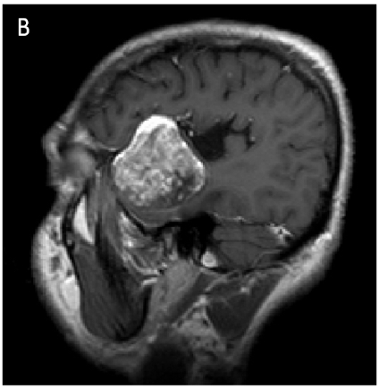
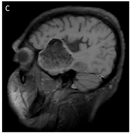
•T1 weighted axial (A) and sagittal (B) image showing an extra-axial hyperintense mass in the left middle cranial fossa. Tiny T1 hyperintense foci are seen in the subarachnoid space along the left convexity(disseminated fat droplets).
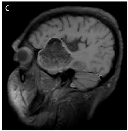
•T1 weighted fat saturated sagittal image (C) showing suppression of hyperintense areas confirming fat content
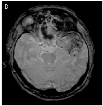
•T2 Gradient echo image (D) showing hypointense areas.
FINAL DIAGNOSIS
• Ruptured dermoid cyst with chemical meningitis.
DIFFERENTIAL DIAGNOSIS
Arachnoid cysts and epidermoid cysts are included in the differential diagnosis of dermoid cysts (4).
CONFIRMATION OF DIAGNOSIS
Presence of fat signal intensity on MRI is always suggestive of Dermoid differentiating it from arachnoid which will show CSF signal intensity and epidermoid which will show diffusion hyperintense lesion with out fat content. The diagnosis may be confirmed by the histopathological examination of the cyst demonstrating the presence of stratified squamous epithelium, sebaceous gland and keratinous debris
TREATMENT
Surgical removal of the cyst with extensive irrigation of the subarachnoid space is the mainstay of management of ruptured dermoid cyst
Ruptured dermoid cyst with chemical meningitis
•Dermoid cysts are thick-walled cysts lined by keratinized squamous epithelium and constitute 0.1% to 0.2% of all intracranial tumors.
•They arise from the inclusion of ectopic embryonic rests of the ectoderm in neural tissue. The cyst material contains skin appendages like hair follicles, hairs, sweat glands, sebaceous glands, teeth or nails (1).
•Dermoid cysts are commonly seen in posterior fossa in midline (2).
•There is a propensity for dermoid cysts to grow into the subarachnoid regions. Dermoid cysts that have not ruptured usually cause only minor symptoms that arise from the tumor’s gradual growth.
•Rupture, which can happen spontaneously or as a result of closed head trauma, is rather uncommon. A cyst rupture that causes aseptic meningitis and the dissemination of fat droplets in the cerebrospinal fluid is the cause of acute symptoms (3).
•Our patient, who had a spontaneous cyst rupture that resulted in chemical meningitis, denied having ever had any trauma-related events.
•The patient’s spontaneous rupture may have resulted from the cyst’s huge size and a weak cyst wall, which lead to dehiscence.
REFERENCES
1.Venkatesh SK, Phadke RV, Trivedi P, Bannerji D. Asymptomatic spontaneous rupture of suprasellar dermoid cyst: A case report. Neurol India 2002;50:480-3
2.Pant I, Joshi SC. Cerebellar intra-axial dermoid cyst: A case of unusual location. Childs Nerv Syst 2008;24:157-9.
3.Rai SP. Ruptured intracranial dermoid cyst. Neurol India 2009;57:98-9
4.Heinze A, Saben R, Hoeschel M. Ruptured intracranial dermoid cyst. Clin Neuroradiol 2008;18:127-8.



Oligodendroglioma
Ganglioglioma, dnet
Dermoid
Left middle cranial fossa dermoid with likely signs of rupture