

Neuroradiology at National Institute of Mental Health & Neurosciences (NIMHANS) has a very long chequered history as old as the institute itself. Hence, tracing the long and illustrious history of neuroradiology requires tracing the history and the objectives and the directives of the institute as the history of neuroradiology is entwined with that of the institute itself closely and that of the dynamic development of neuroradiology in the rest of the world at large. The department of Neuroradiology at NIMHANS has been a pioneer in the development of the specialty of neuroradiology in the country.
What started as a Lunatic Asylum in the later part of 19th century was later transformed into a Mental Hospital in Bangalore in the early part of the 20th century. In the year 1925, the name of the Institution was changed from Lunatic Asylum to that of Mental Hospital under the vision of the then Maharaja of Kingdom of Mysore. The Mental Hospital was shifted in November 1937 to the present location. Under the recommendation of Bhore Committee in the year 1946, the Government of India felt the need for training personnel in the various fields of mental health and ancillary specialties for the country and the Mental Health Institution was reorganised. The All India Institute of Mental Health was thus established in 1954 under the joint funds of Government of India and Government of Karnataka.
The Neuroradiology Department was established in the year 1958 along with the departments of Neurology and Neurosurgery. This was hence the first department of neuroradiology established in the country. This was headed by Dr. K. Mahadevan Pillai, who was appointed as Associate Professor of Neuroradiology. The Department was started in an extended room attached to one of the pavilions of the Psychiatric Wards. This room was specially designed for the purpose of radiological work. Dr.K.M.Pillai was trained in neuroradiological procedures and was able to do specialised techniques like percutaneous angiography, air and positive contrast ventriculography, etc. He continued the work as an Associate professor until September 1962, In 1973, Dr. N.C.Gupta joined as Assistant Professor of Neuroradiology on deputation.
NIMHANS was established on 27th Dec 1974 under the orders of the President of India. The three point charter of NIMHANS included service, manpower development and research. The main ethos of the institute being a multidisciplinary team approach for promoting preventive and curative aspects of mental health and the interdepartmental integration as the mainstay of the Institute. Under the guidance of Dr. R.M.Verma a full-fledged neurocentre was established. Neuroradiology was a central part of this neurocentre placed in the ground floor of this new building. The department took a more formal footing under the guidance of a young dynamic radiologist, Dr. N.C. Gupta. He continued until April 1976 when he was transferred to Goa. In the year 1977, Dr. B.Vasundhara Devi, a radiologist was appointed as a Lecturer in Neuroradiology and she was in-charge of the department. Subsequently, in the year 1979, the Department was further strengthened by the appointment of Dr. B.Y.T.Arya, who joined as senior resident and later on became Additional Professor and the Head of the Department.
The Department at that time in the late seventies was equipped with 1000 mA routine X-ray equipment, ‘Pleophos’ with Potter-Bucky (Gigantos) , 500 mA Elma Schonander skull table,, ‘Klinoscop’ 500 mAs with Image Intensifier for Myelograms and a 60 KV portable X-Ray Unit. The 1000 mA X-Ray unit ‘Gigantos’ was also used for taking radiograph of skull and stereoscopic surgery in the operation theatre. The radiographs could be taken from outside the OT through a glass window. A ‘Cordis’ Pressure Injector was added in 1980, a automatic processor in 1981, a Planigraphic Tomogram table in 1982 and a fibre-optic image intensifier was attached to the Klinoskop table in 1983 which facilitated a better and clearer visualization of the structures on fluoroscopy. The radiographs taken are still being utilised for various teaching purposes.
A paradigm shift in the sophistication of the services of the department happened with the installation of the first CT scanner in NIMHANS in 1982. This was a second generation ‘Head scanner’. This was only the second generation scanner in the country. With the introduction of the C.T. Scanner, a number of invasive investigative procedures like ventriculogram, pneumoencephalogram etc., became obsolete and procedures like angiograms reduced. Oil- contrast myelograms and epidurograms continued to be practiced.
A second reason for the shift was the installation of a ‘Biplane Angiography’ equipment in 1984. This started the era of transfemoral catheter angiography of the cerebral and spinal vessels, with considerable reduction of radiation to the patients and operator. This was soon to prove to be a major leap in the services of the department as interventional procedures started soon thereafter. This was largely due to the visit and demonstration of these techniques by none other than Prof. Luc Picard, from Hospital St. Julian, Nancy, France in 1985. His visit spurred the young faculty in the department to pursue the field of interventional Neuroradiology further by acquiring additional skills and knowledge. With this agenda, two members of the faculty, Dr. BYT Arya and Dr. PN Jayakumar worked in his department as fellows for period of a year. With this additional training, therapeutic neuroradiology came to be established as a routine part of treatment of neurological disorders.
The last decade of the previous century was a breakthrough for the neuroradiology department. The department grew exponentially in terms of services, manpower development and research. This was due to many factors including acquiring new equipment, starting new courses, increasing patient services and novel research enterprises.
Profile of NI&IR 2000-2010
Over the years it developed into a internationally known centre for both clinical services & research activities. In keeping with the functional profile, the department was renamed as Department of Neuroimaging & Interventional Radiology.
Clinical services:
The department provided both diagnostic and therapeutic services. The diagnostic services include, CT scan imaging services to cater to the patients in the OPD & the casualty. A CT scanner was housed in the casualty & OPD block to take care of emergency requirements round –the-clock. In addition, X-ray services were also provided in the outpatient and casualty round –the- clock. An additional CT scanner provided state- of- the- art diagnostic capability and was located in the neurocentre. State- of- the- art MRI scanner provided for the best of neuroimaging capabilities in this part of the country. Therapeutic neuro-radiological procedures became routine practice and included procedures like endovascular treatment of intracranial aneurysms, Arteriovenous malformations , Arteriovenous Fistulae and tumors, carotid and vertebral angioplasty, intra-arterial and intravenous thrombolysis in patients with arterial & venous stroke. This was largely due to the fact, that the faculty were trained in advanced techniques of neurointervention at internationally acclaimed centres of excellence. Dr. PN Jayakumar was trained by Dr. Luc Picard and Dr. Jacques Moret, France; Dr. MK Vasudev was trained by Dr. Guisippe Scotti, Italy and Dr. SG Srikanth by Dr. Anil Gholkar, UK .
Equipments:
The diagnostic armamentarium in the department was always state- of-the- art imaging equipments. A 1.5 Tesla MRI system ‘vision’ provided routine MRI examination and also functional MRI, MR spectroscopy, MR Angiography. Biplane Digital subtraction Angiography ‘Neurostar’ with facilities for ‘rotational Angiography’ and 3-D Angiography delineated the pathophysiology, angiographic anatomy and blood circulation and in addition supported the interventional procedures in the treatment of vascular lesions. The latest additions of multislice spiral CT Scan ‘Volume zoom’ gave more thrust to the high techniques of care like high resolution ultrafast imaging, CT Perfusion, 3D CT scanning, virtual endoscopy and stereotactic biopsy procedures.
Man Power Development
The faculty was chaired by Prof. PN Jayakumar initially followed by Dr. MK Vasudev as Addl. Professor, Dr. SG Srikanth as Associate professor and Dr. Jerry ME Kovoor as Assistant Professor. The end of this period was marked by the recruitment of Dr. Rose Dawn Bharath and Dr. Arvinda HR as Assistant professors after completion of DM in Neuroradiology from NIMHANS and SCITMST respectively. Senior residents form the rest of the clinical staff. The services are supported by a technical staff of fifteen qualified technicians trained in all aspects of patient examination in all the modalities, staff nurses of eight and administrative staff of three.
The department was continuously engaged in various teaching programmes and academic research activities with the view of establishing excellence in the subject of Neuroradiology. A national committee consisting of pioneers in the fields of radiology emphasized the need to establish a superspeciality course in neuroradiology. The department was recognized as the center to start the DM course programme in neuroradiology. The department started a three year post-MD programme leading to DM degree in neuroradiology since year 2000. Two DM neuroradiology students were admitted in each academic year with a total strength of six senior residents and two junior residents undergoing training in all aspects of neuroradiology. Post graduate students of diagnostic radiology from other medical colleges of the state and country were routinely trained in neuroradiology. In addition, post-graduate students of DM neurology and MCh neurosurgery were posted to the department during their course. The department also started a one year post-Diploma course in neuroradiology for radiology technicians during this time.
The developments in the department of NIIR in the last decade was many folds not only in the addition of newer state- of- the- art equipments like MRI, Spiral CT scan, DSA etc but also in the academic activities. The department hosted many national conferences & workshops on neuroradiology and neurointervention attended by many national & international faculty. The department hosted annual programme on advanced techniques in neuroimaging. The faculty members were involved in many national & international projects.
Research
The department continued to be in the forefront in research in neuroradiology in the country because of which it attained a status of eminence. The department was engaged in clinical research in various aspects of neuroimaging. The research projects were funded by various agencies like NIMHANS, NGOs, national and International organisations. The department as a part of the institute attained international standard and entered into collaborative research projects with major centers in USA, in neurological and psychiatric illnesses.
Profile of NI & IR 2010 – 2021
Equipments
The period 2011 to 2020 unraveled the metamorphosis of neuroimaging with the focus of patient care, teaching and research expanding the frontiers to include structural neuroimaging to functional neuroimaging and then to molecular neuroimaging. This was reinforced by the acquisition of additional equipments a 1.5 T MRI Aera (Siemens), 3T Ingenia (Philips), 3T Skyra (Siemens- Project), 3T MRI-PET (Biograph mMR: Siemens), 1.5T Intraoperative MRI( Optima MRI 450W), 16 slice CT scanner in the Neurocenter for inpatients and minimally invasive procedures. Digital radiography unit and amplified portable imaging with portable x-ray in each floor further improved the availability of imaging in critically ill patients. Genesis of the emergency wing of NI&IR in the year 2008, with a 16- slice CT scanner Brilliance (Philips) and a digital Radiography unit with recruitment of technologists under contract to manage this unit while awaiting sanction of new posts was another major development. This unit was augmented with the addition of a second 16 slice CT Unit (Bright Speed GE) in the year 2012. In the year 2019, with the purchase of a 64 slice CT scanner (GoTop Siemens) all equipments in the emergency wing were brought under one roof making the process very efficient. A fully functional PACS (Meddiff upgraded to Centricity PACS) with the required hardware for storage (GE) was installed with a disaster recovery unit which lead to initiation of online reporting. The field of neurointervention, also revealed exponential growth during this time with acquisition of the second Biplane DSA (Allura clarity Philips) and initiation of acute intervention program in stroke. These equipments and focused skill set augmented patient care with 15- 20% annual growth in referrals for CT, MRI, DSA and USG. By the end of 2020 we were witnessing around 2500 diagnostic and Intervention cases in that year.
The second decade of the 21st century marked the birth of Molecular Neuroimaging and Acute stroke intervention programs in the department of NIIR.
Molecular Neuroimaging Center
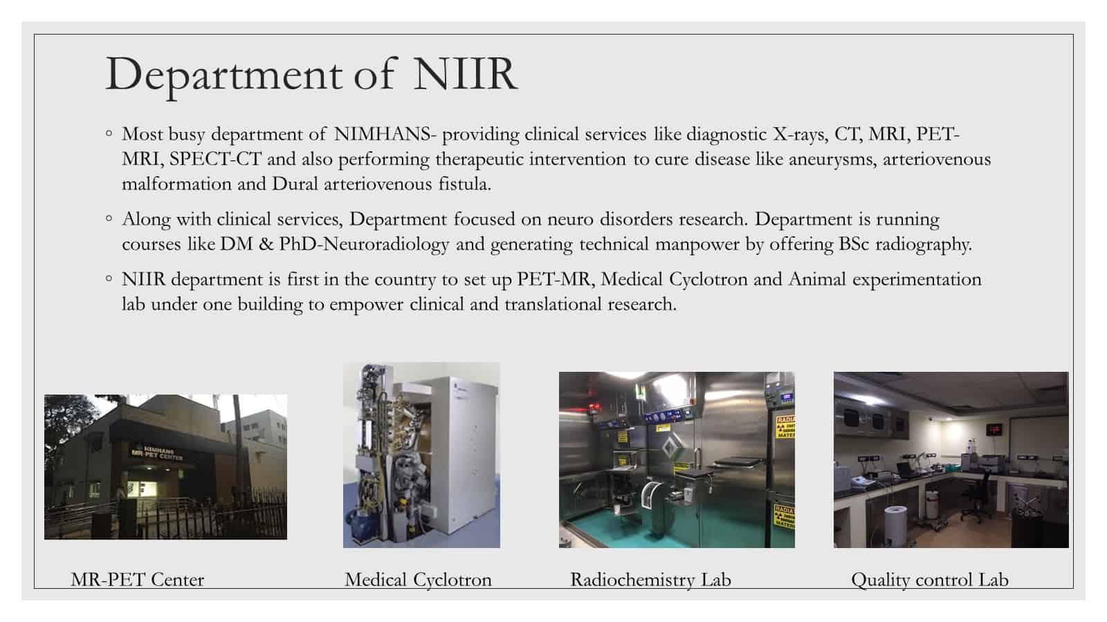
- Inauguration of the MRPET- Cyclotron facility at NIMHANS in 2015 by the Honourable Prime Minister Shri Narendra Modi marked the beginning of the field of Molecular Neuroimaging at NIMHANS and initiated patient care from April 2015.
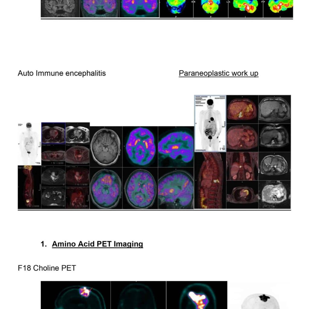
- Within 3 years MR PET imaging became a standard of care for several neurological disorders. We started diagnostic work-up packages for diseases such as epilepsy, dementia, autoimmune encephalitis, paraneoplasm and metastases. These were also used for several patients to assess response to treatment and for follow up.
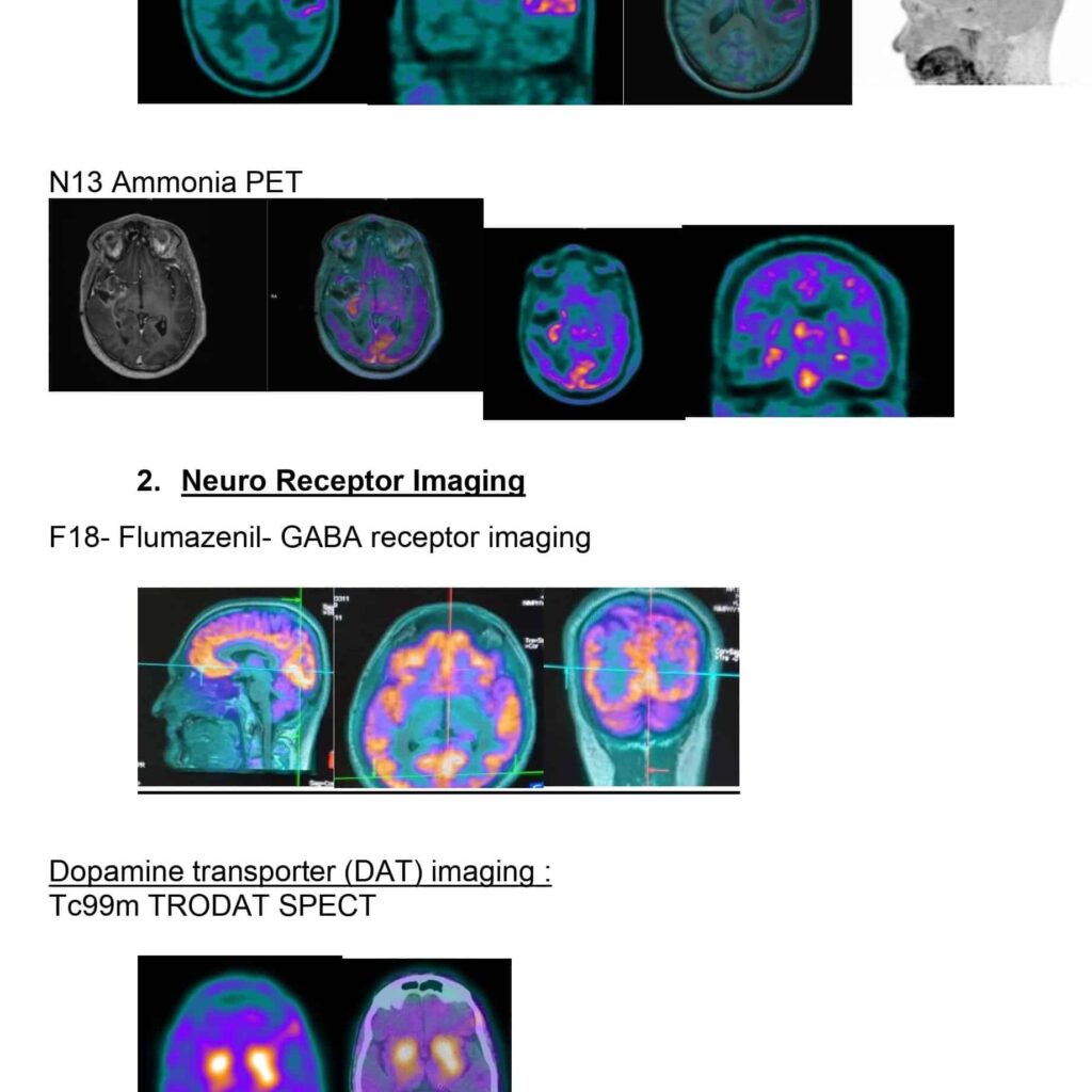
- In 2018, with the licensing of Medical Cyclotron, we became self-reliant and started in-house production of all radiopharmaceuticals for clinical use. Apart from F18 based glucose radiotracers we also started synthesis of amino acid radiopharmaceuticals and neuroreceptor based radiotracers for patient care for the first time in the country.
- Amino acid imaging tagging methionine, choline and tyrosine added more precision to tumor imaging and could change the management strategy in several patients.
- Dopamine imaging using TRODAT or FDOPA has become a crucial diagnostic tool in patients with Parkinson’s disease allowing higher confidence in their diagnosis.
- In 2020, within 5 years of its inception, we initiated commercial synthesis of radiopharmaceuticals, extending our services to patients beyond the boundaries of NIMHANS and began the process of generating revenue for the institute with a vision to be self reliant.
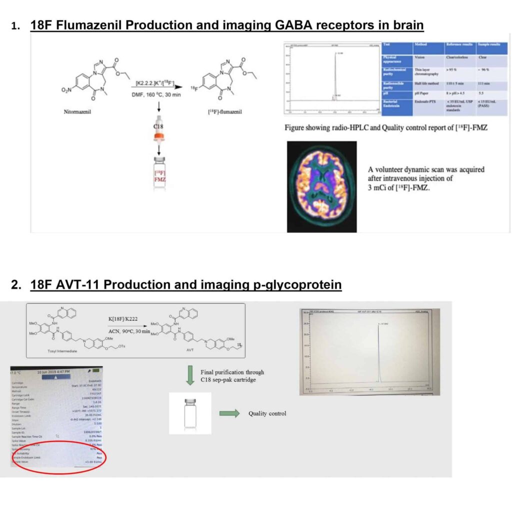
- In the field of research, in vivo imaging of GABA receptors in human brain and with the synthesis of 18F- AVT11 imaging p-glycoprotein expression was initiated with the synthesis of F18 Flumazenil.
- This centre could attract funding from several scientists and PhD students and we are currently working on translational research in high grade gliomas using artificial intelligence based methods for predicting survival and using stem cell research in understanding chemoresistance in glioblastoma
- Molecular Neuroimaging in Patient care
- F18 FDG imaging
Acute Ischemic Stroke
Indian scenario:
Acute ischemic stroke (AIS) is a leading cause of disability and morbidity. In a developing nation like India ,it assumes a much greater significance . In addition AIS due to large vessel occlusion (LVO) is significant cause of mortality .Unfortunately data regarding the epidemiology of AIS in India is scarce. As per a systematic review ,prevalence of AIS is around 44.29-559/100000 per year in different parts of the country. As per a stroke registry ,data collected in 2005 showed an age adjusted cumulative incidence per 100000 people to be 1,174 cases in Bengaluru, this being higher than other parts of the country.
NIMHANS advantage:
Being one of its kind quarternary referral centres for neurological diseases, NIMHANS has strategic advantages to it when compared to any other centre:
1) Centrally located with good connectivity
2) Only centre in the country to have a casualty dedicated to neurological emergencies managed by emergency neurology, neurosurgery and neuroimaging and interventional radiology teams on call.
3) Dedicated emergency imaging wing of dept of NIIR with two CT scans 128 slice CT, 16 slice CT.
4) Two biplane equipped dedicated neuro-angio suites
5) Round the clock advanced MRI facility
July 2018: Beginning of acute ischemic stroke intervention program at NIIR:
Acute ischemic stroke intervention program was envisioned and brought into inception in July 2018. An acute stroke team was formed with a Interventional neuroradiology faculty, neurologist , senior residents from both departments ,with support from neuro anesthesia and neurosurgery departments .An AIS workflow was established with incorporation of nursing staff ,radiographers.
First mechanical thrombectomy was done on 8th July 2018, with successful complete recanalization of left Middle cerebral artery occlusion. Following this, till date 125 mechanical thrombectomies have been done by the end of 2020.
NIIR acute ischemic stroke workflow:
All acute ischemic stroke calls are attended by duty neuroradiologist with immediate interpretation of the imaging and Cathlab team is informed .Patient selection is done as a team with neurology, there is simultaneous preparation of the angio suite for possible procedure and neuroanesthetist is informed by NIIR resident. Once the patient is deemed eligible for thrombectomy, he/she is shifted to a already prepped cathlab. Post intervention patient is shifted to ICU, ward depending on the clinical status. Post procedure care is done by NIIR and neurology team together.
Hallmarks / strengths of NIMHANS NIIR stroke intervention program:
1) Interpretation of neuroimaging including immediate post processing of advanced imaging like perfusion in cases of wake up stroke done by dedicated neuroradiologist increasing the diagnostic accuracy when compared to interpretation by general radiology or emergency medicine teams at other centres.
2) Biplane neuroangio suites – improved resolution and decreased procedure time. Xper CT facility allows on table cone beam CT when needed.
3) Six-bedded observation room is attached to the angiosuite. It has a patient: nurse ratio of 2:1. IVT has been done here with better dedicated continuous monitoring and patients taken for rescue mechanical thrombectomy.
4) Mechanical thrombectomy technique used is ADAPT. The current program has by far one of the highest numbers of ADAPT procedures done in a single year
5) A novel selection method for wake up stroke is being used in few cases with NIHSS-CTA aspect mismatch. This method has real world applications where in delays in time to recanalization are much more than in trial.
6) A real world approach with patient benefit being foremost priority, even in its nascent stage the program also included a select few patients over 80 year of age, with ASPECT scores of 5 for mechanical thrombectomy with good outcomes. Tandem lesions, rescue intracranial stenting also has been done in few of the cases.
7) Approval of Karnataka Arogya Scheme was given for mechanical thrombectomy procedure, a game changer which has allowed the dept to extend the benefit to patient who couldn’t afford this procedure.
8) STROKE AUDIT: Each and every MT case is discussed in the audit meet attended by departments of neurology and NIIR. This has led to continued improvement in workflow with significant reduction in door to recanalization time.
Limitations of the program.
1) Dedicated ICU bed unavailable for most of the patients post procedure.
2) Cardiologist for management of patients with cardiac morbidities like AF, LVF
3) Material cost
VISION
A) Dedicated stroke ICU/ beds (02) for patients undergoing procedure.
B) Upscaling of the number of procedures:
Increasing patient awareness and education – posters in metros, buses, education apps
Collaboration with nursing homes, hospitals with lack of facility to provide mechanical thrombectomy to develop –“drip-ship” and “mothership” model of workflow for efficient referral.
Decreasing material costs- Corporate funding, NIMHANS Health /Stroke card (patient funded low cost health insurance like model)
With recent metanalysis establishing benefits of MT even in ASPECT <5, the patient inclusion can be expanded if dedicated ICU facility is present.
C) Tele- stroke -Hub and spoke model:
Development of web portal with hospitals within this model for providing expert services for neuroimaging interpretation and IV thrombolysis.
D) Participation as site for new device, revascularization trials
E) Creation of AIS registry
F) Collaboration with government cardiac institutes of excellence for locum cardiologist for the program.
G) Development of deep learning prediction model based on the registry data for identification of factors affecting outcomes of revascularisation in Indian scenario.
Human Resource Development
In Nov 2011, Prof. AK Gupta joined the department as head of the department after retiring from Sree ChitraTirunal Institute of Medical Sciences. Subsequently, Dr. Hima Pendharkar, Dr. Jitender Saini, Dr. Chandrajit Prasad, Dr Maya D Bhat joined the team as assistant professors after completing their DM in Neuroradiology. In the year 2015 with the initiation of molecular neuroimaging wing, a multispecialty team with Dr.Sandhya M, Dr. Chandana N and Dr.Pardeep Kumar joined the team as faculty along with 3 group A technical team. With focus on function preserving therapies, Dr. Karthik K and to initiate translational Neuroimaging, Dr. Manoj Kumar joined the team of faculty. The technical team also saw recruitment of 7 more new staff with upto 50% replacement of the staff strength.
Research
Research also witnessed prodigious growth with change of philosophy from image acquisition to image processing. The foundation of this philosophical change was laid during the visit of Prof Bharat Biswal and the team in the year 2012.Subsequently data driven methods empowered the faculty and students in answering several clinical questions using imaging methods resulting in publication of several research papers as prime authors in high impact journals. Faculty of NI&IR also initiated several interdepartmental and inter-institute collaborative projects using these methods. Initiation of Dr. Mahadevan Pillai Memorial teaching programs for radiology students, conduct of hands on training workshops in functional MRI at CMC Vellore, PGI Chandigarh and the conduct of the largest national level meeting of the ISNR exemplified the training philosophy during this period.
Broad areas of research activities include
Dementia: Structural and functional connectivity in Alzheimer’s dementia, Fronto temporal dementia etc. Dr Sandhya M is the faculty who is interested in this field.
Neuropsychiatric disorders: More than 30 imaging based projects are currently underway using the techniques of fMRI and diffusion weighted imaging in the field of OCD, addiction and schizophrenia. Dr. Rose dawn Bharath, Dr. Jitender Saini and Dr Chandrajit Prasad has several collaborative projects with faculty from department of Psychiatry.
Cognition and language: Neurohemodynamic correlates of cognitive networks and the effect of education and normative multilingualism on these networks is being studied using task based fMRI, resting state fMRI and fMRI EEG. Dr. Rose Dawn Bharath is the faculty who is interested in this field
Neurometabolic disease: Application of newer MR imaging techniques for the diagnosis of neurometabolic diseases. Creation of large MRI database of metabolic especially Wilsons disease and mitochondrial cytopathies. Dr Chandrajit Prasad and Dr Maya D Bhat has several collaborative projects with the department of Neurology
Movement disorders: Investigation in various movement disorders have been carried out. Large sample of spinocerebellar ataxia have been studied previously and aim to carry out further research in these disorders especially on SCA1, 2, 3 and 12. Similar studies in PSP patients with aim to characterize subtypes of PSP using advanced MR techniques. Resting state and motor functional connectivity in writers cramp and its modulations using rTMS has been assessed. Dr. Rose Dawn Bharath and Dr. Jitender Saini has several research projects in this field
Neuroinfections: Serial MR Perfusion markers were used to derive biomarkers that can predict response to treatment. Dr A K Gupta and Dr Rose Dawn Bharath guide students in these topics.
Epilepsy: The institute has a dedicated epilepsy team and quantitative imaging studies are being conducted on various epileptic disorders like focal epilepsies, Juvenile myoclonic epilepsy and reflex epilepsy especially hot water epilepsy. Imaging contrasts like diffusion tensor imaging and functional MRI are being used to study these disorders. EEG f-MRI application in various seizure disorders is another area of research. Dr Rose Dawn Bharath and Dr Jitender Saini has several research projects in this field
Brain tumors: Brain tumors is another area where research imaging projects are being carried out. In the past studies have been done on proton and phosphorus spectroscopy for characterization of the brain tumors. Current areas of interest are perfusion and diffusion weighted imaging for characterization of various brain tumors. Efforts are being made to study newer perfusion imaging techniques like DCE for characterization of brain tumors. Dr. Rose Dawn Bharath currently works on peritumoral imaging genetic correlation and texture based glioma grading, cortical plasticity following glioma surgery, Dr. Jitender Saini works on ASL perfusion in grading of glioma and Dr. Chandrajit Prasad works on segmentation approaches on glioma grading.
Demyelinating disorders: Focus on various aspects of acquired inflammatory disorders like multiple sclerosis, neuromyelitis optica, ADEM etc is carried out in collaboration with the department of Neurology. Dr Rose Dawn Bharath, Dr Maya D Bhat guides students in these fields.
Traumatic brain injury: Research work has commenced in this direction during last few years. In a recently concluded study longitudinal changes in the brain morphology and connectivity using DTI have been studied. Dr. Rose Dawn Bharath and Dr. Jitender Saini has several research projects in this field
Anesthesia and pain: Small pilot projects have been carried out in the field of anaesthesia and pain imaging. Dr. Jitender Saini and Dr Arvinda H R has collaborative projects in this field
Interventional neuroradiology: Research areas include Idiopathic intracranial hypertension, management of direct CCF and spinal vascular malformations. Dr AK Gupta, Dr Hima S Pendharkar, Dr. Arvinda HR and Dr Chandrajit Prasad guides several students in these topics.
Molecular neuroimaging: Research on development of newer molecular imaging markers and applications as elaborated above are being conducted. Faculty interested in this program is Dr. Rose Dawn Bharath, Dr. Sandhya M, Dr. Chandana N and Dr Pardeep K
Translational Neuroimaging: Dr. Pardeep Kumar and Dr. Manoj Kumar have initiated translational imaging with in vitro imaging in glioma cell lines.
Academic Program
DM Neuroradiology course was renamed as DM in Neuroimaging and Neurointerventional Radiology, and given the popularity of the course; the number of seats was increased to four students per year. Structured teaching program expanded to functional and molecular neuroimaging and hands on interventional neuroradiology. Log book including the spectrum of cases they have managed and also indicating the number of interventions performed independently by the candidates were reviewed periodically. Faculty coordinators were selected to be mentors for these students. Students after completing the program were absorbed at reputed neuroradiology centers both in India and abroad.
BSC Radiography course also continued with intake of 10 students per year. Internship program was initiated to provide hands on comprehensive training in all modalities. Several students were selected as application specialists, some went abroad and many started working in high volume imaging centers.
PhD Neuroimaging and Interventional radiology program became more popular and graduate engineering students joined the program and are currently working on translational neuroimaging using machine learning and molecular imaging markers. The state of the art animal MR Imaging centre is expected to add value to this program. Several established researchers of international repute migrated to the institute through national fellowship schemes such as Ramalingaswamy fellowship.
The number of students visiting NIIR for short term training also increased in number spanning radiology, neurology, neurosurgery, psychiatry fields. They came from several parts of the state and across the country to learn from the rich plethora of patients visiting the institute. Besides, this period also witnessed the faculty and students proactively forming interdisciplinary subspecialty groups for epilepsy, movement disorders, tumors, vascular diseases, neurodegenerative disorders and dementia.
The department of NIIR has over the course of last two decades strived to be successful in setting exemplary standards in clinical services and research across all domains of mental health and neurosciences both in India and this part of the world. The department aims to see beyond the boundaries of clinical medicine and is now exploring the use of translational neuroimaging methods in understanding disease neurobiology and clinical outcome.
Dr. PN Jayakumar
Retd. Prof. & Head
Dr. Rose Dawn B
Prof. & Head.